Ex vivo small bowel auto-transplant
Whipple with sub-total colectomy with SMV and SMA reconstruction
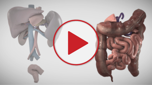
View the Video
Video Chapters
Case Description
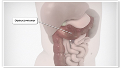
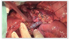
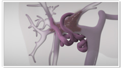
- In this video we present a technical challenge that required both surgical oncology and transplantation techniques to overcome. The patient is a 25 year old man with Lynch syndrome who developed an obstructing tumour in the mid-transverse colon. The tumour eroded posteriorly into the pancreas, obliterating the distal SMV and abutting the SMA.
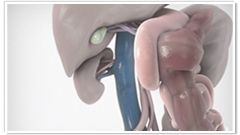
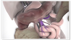
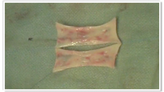
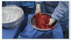
- After a decompressing ileostomy, the patient had multiple courses of chemotherapy with mild response of the primary and no metastases becoming apparent. It was elected to being him to the OR for a subtotal colectomy with whipple, reconstructing the SMV (proximal bifurcation and first jejunal branch) to a panel graft created from the superficial femoral vein.
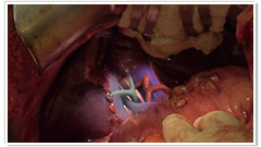
- The final dissection and venous reconstruction were performed ex vivo after flushing the intestines (small, large) and pancreas head with ice cold preservation solution on the backbench. After re-implantation, the splenic vein was anastomosed to the side of the panel graft in order to avoid splenic congestion.
- The patient did well post-operatively, and was discharged home on post-operative day 10. His 3 month follow up CT shows patent vessels. This case demonstrates the marriage of transplantation techniques to hepatobiliary/surgical oncology procedures.
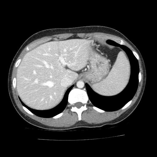
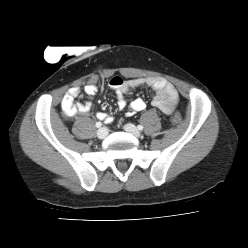
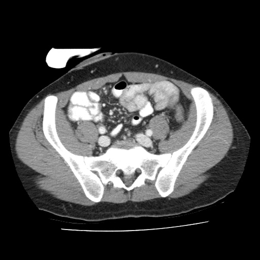
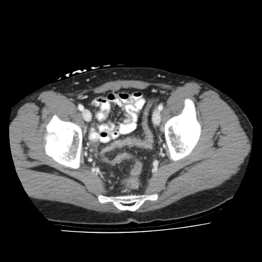
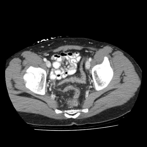
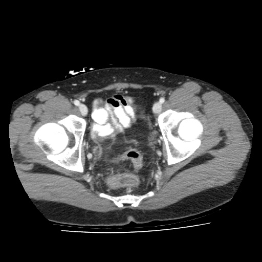
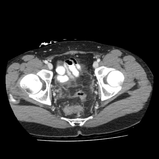
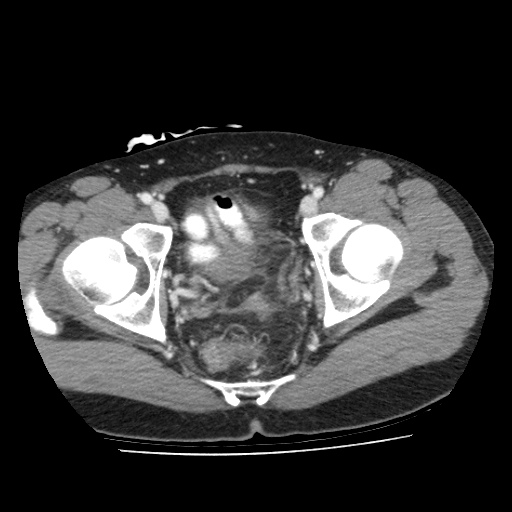
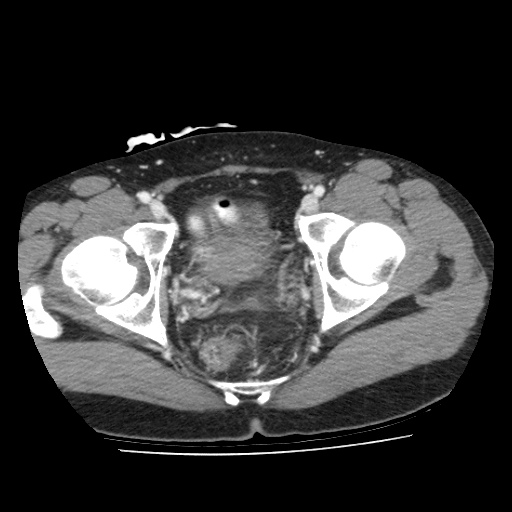
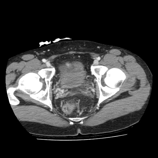
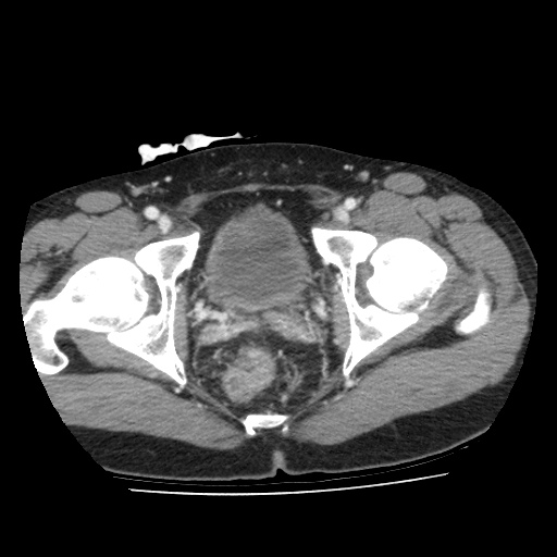
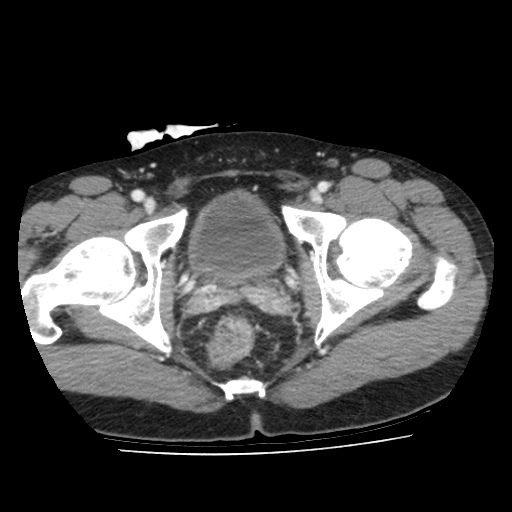
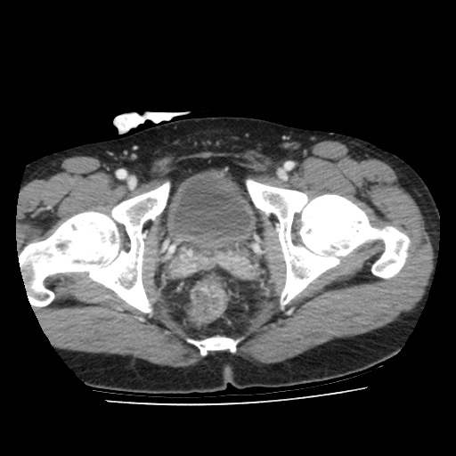
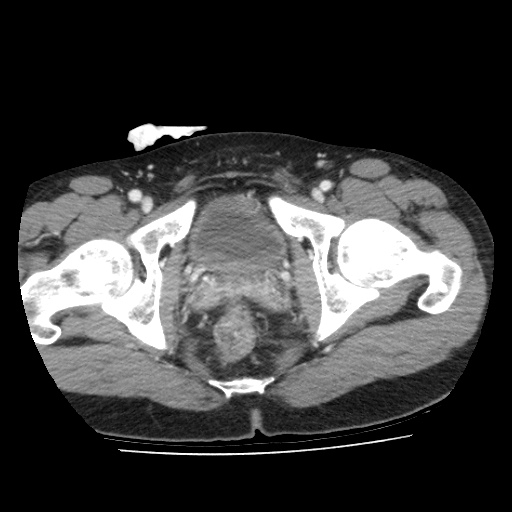
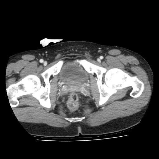
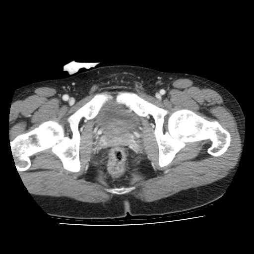
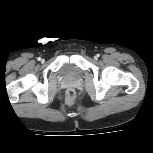
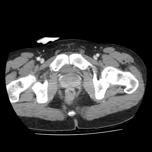
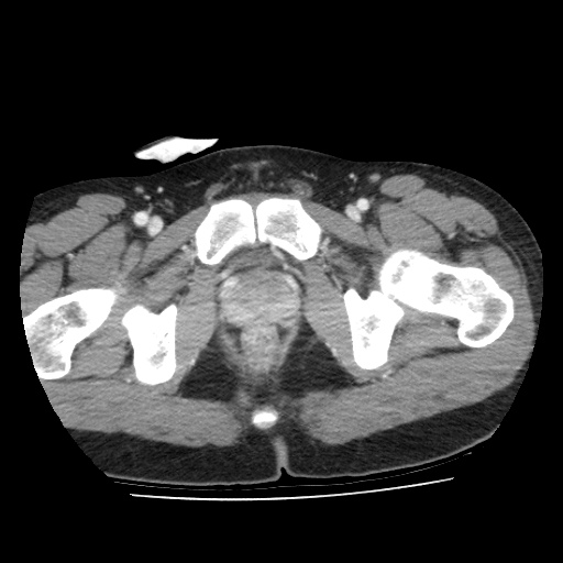
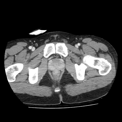
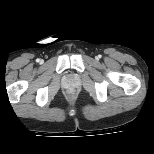
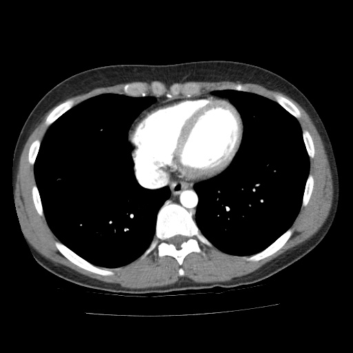
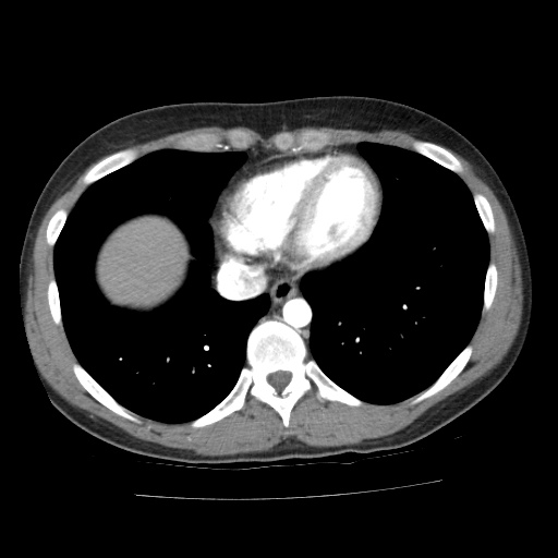
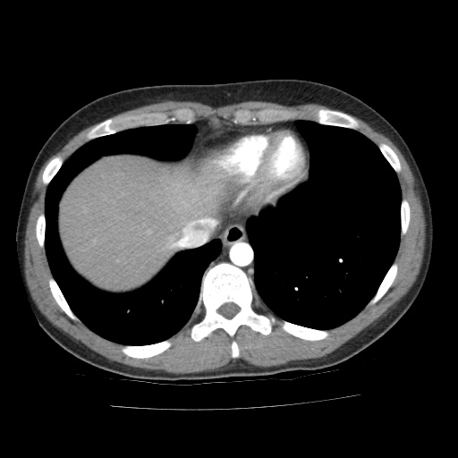
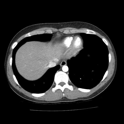
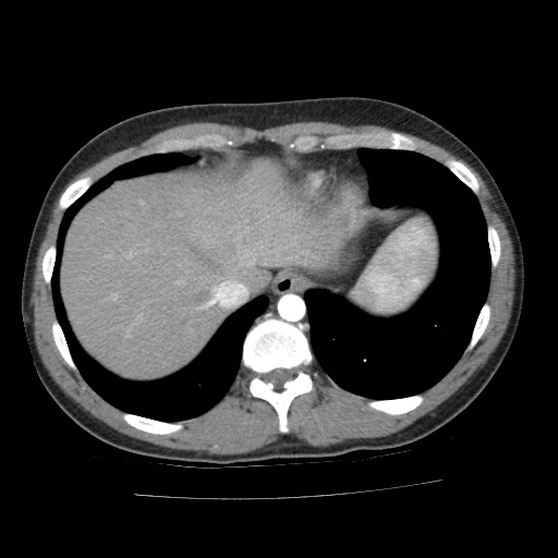
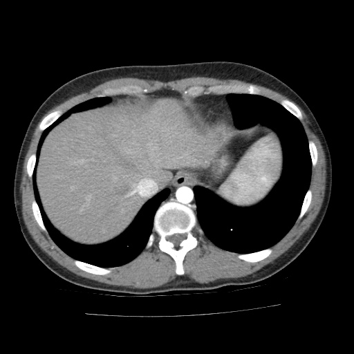
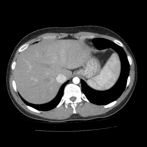
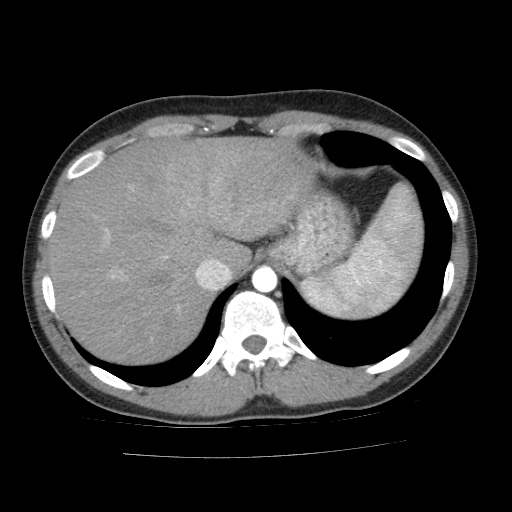
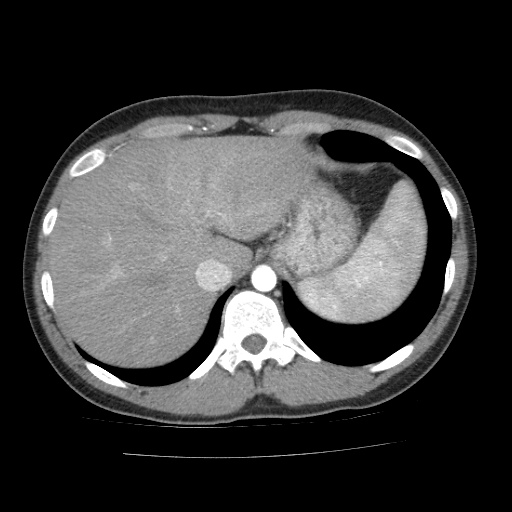
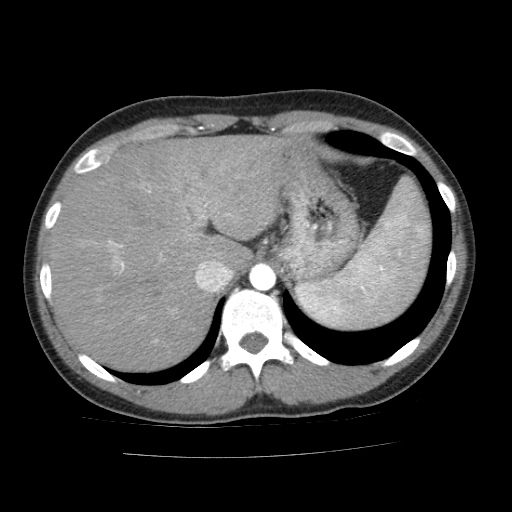
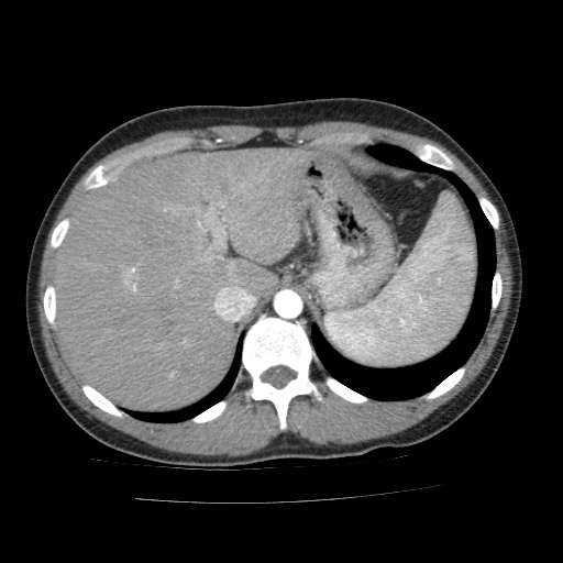
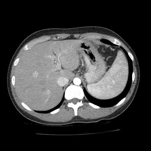
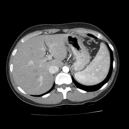
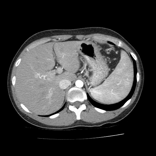
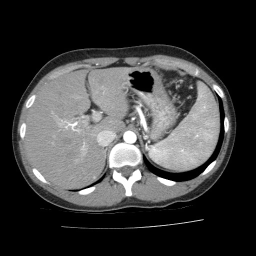
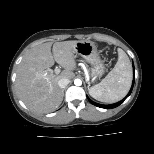
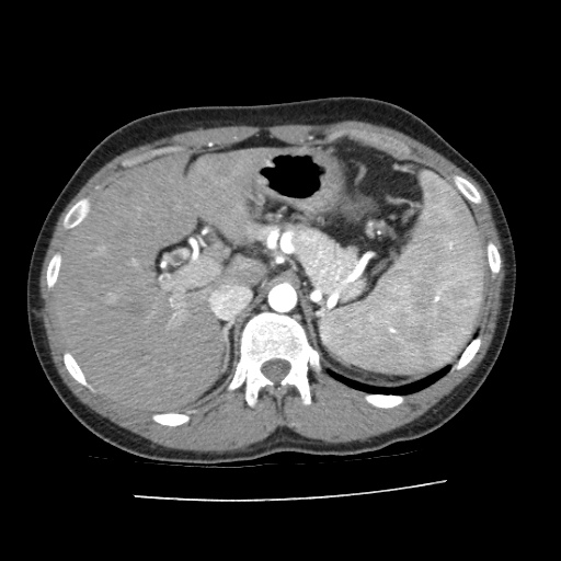
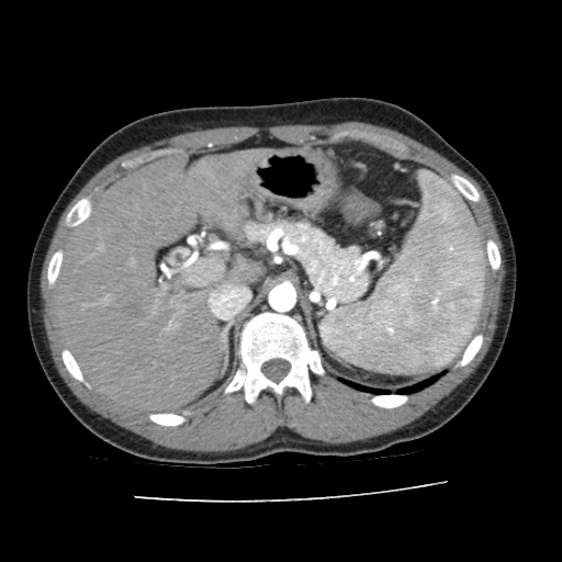
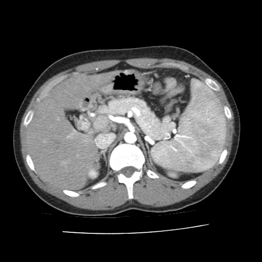
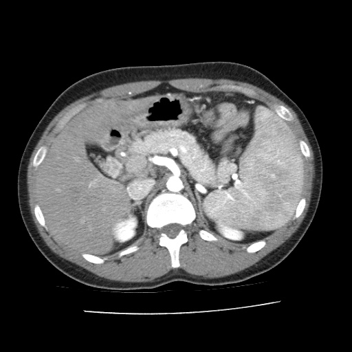
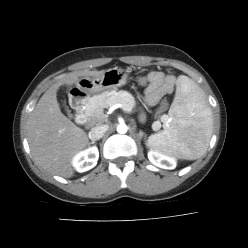
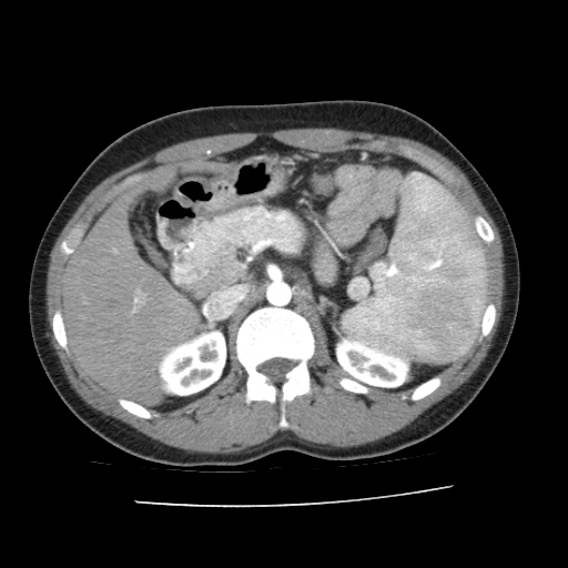
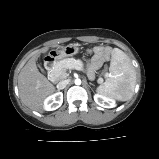
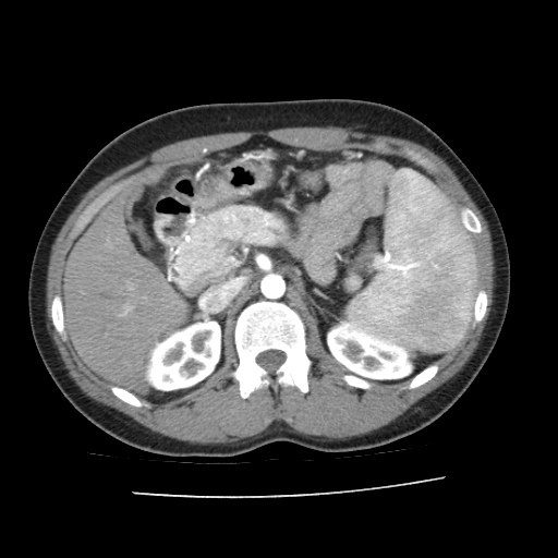
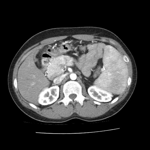
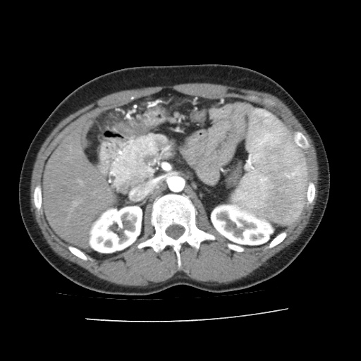
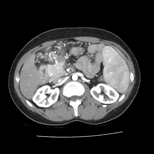
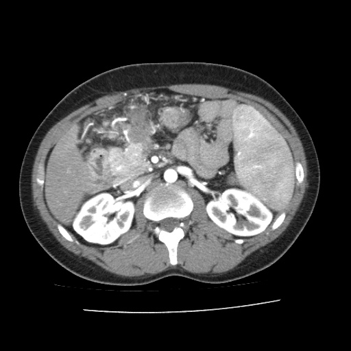
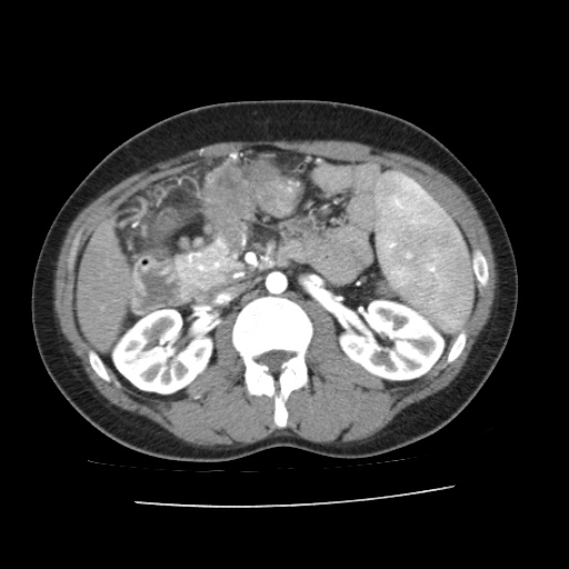
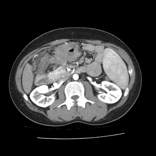
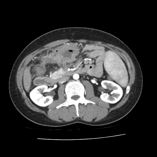
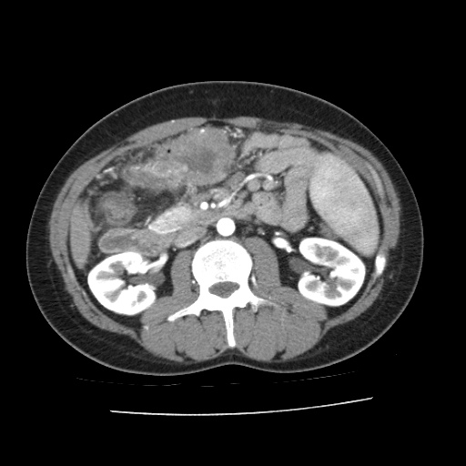
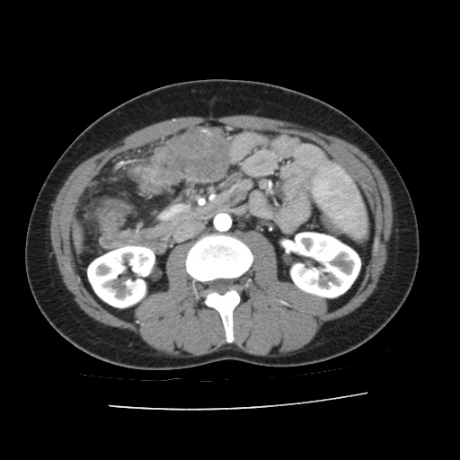
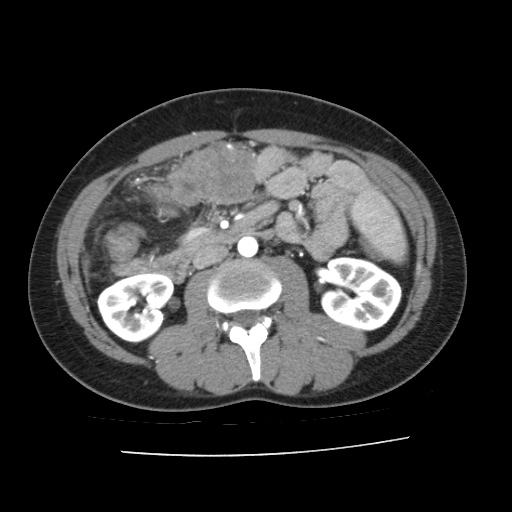
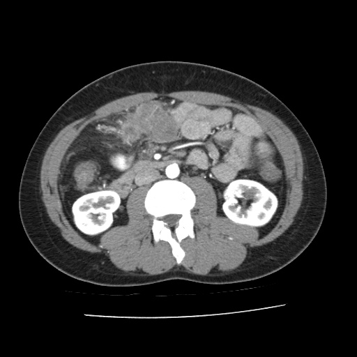
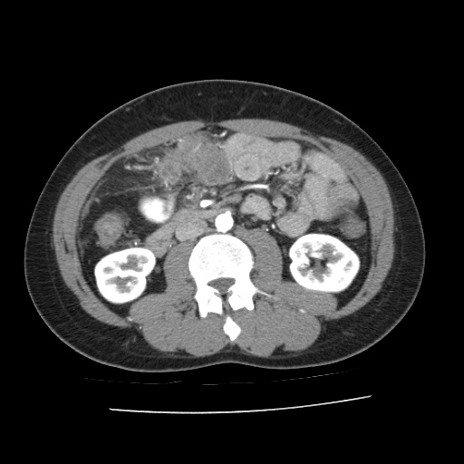
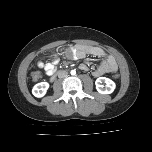
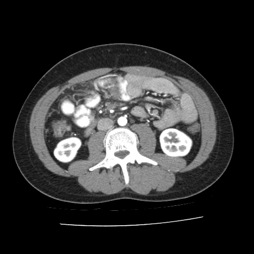
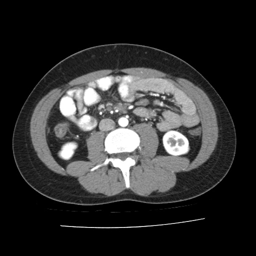
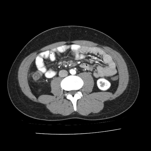
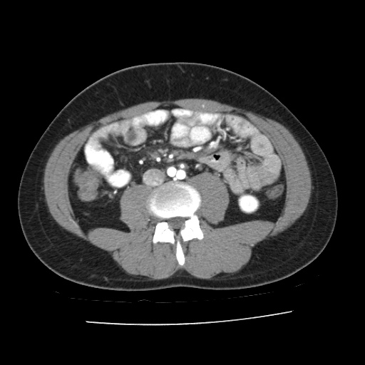
CT scans (venous phase)
- Click and drag the slider on the left to transverse through the venous CT series. Clicking on the grey button on the bottom will reveal clinical findings by the surgical team; click on the descriptions to bring the image up to the main window. Best viewed in Mozilla Firefox, Google Chrome or Safari.
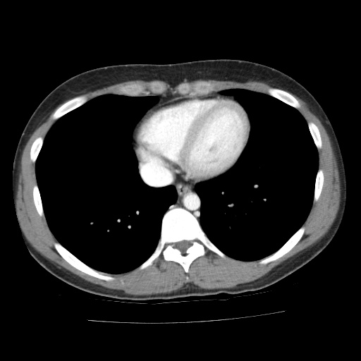
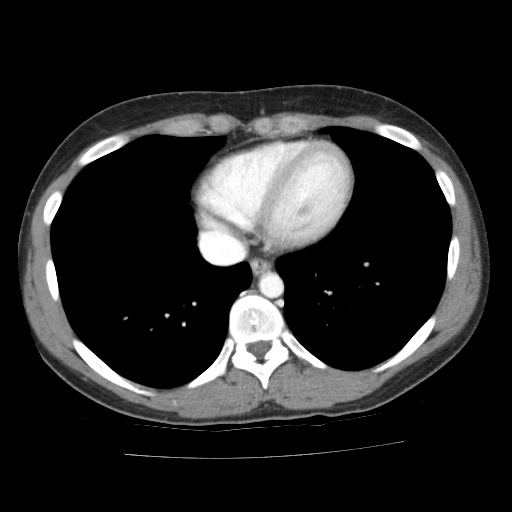
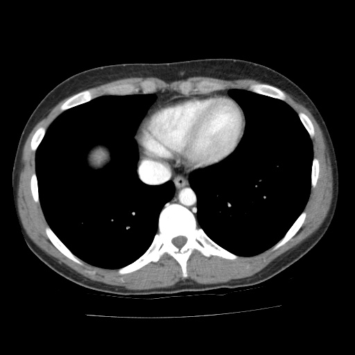
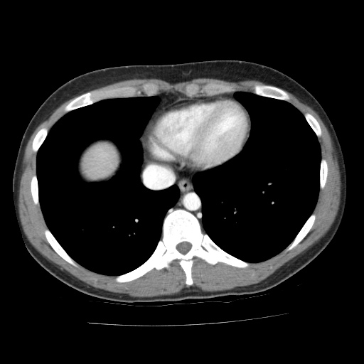
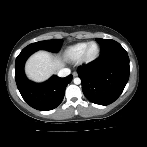

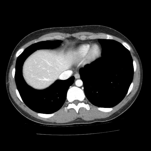
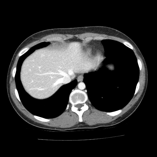
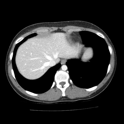
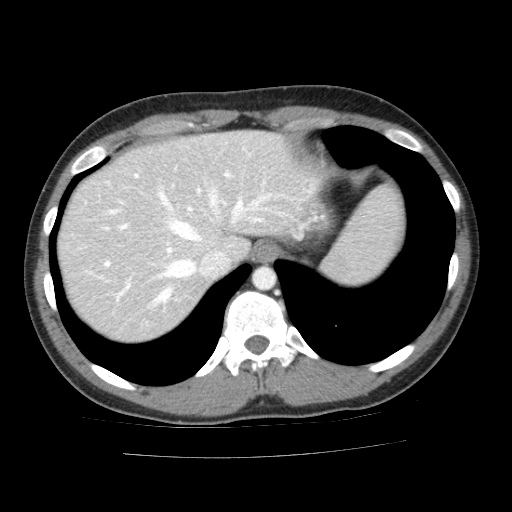
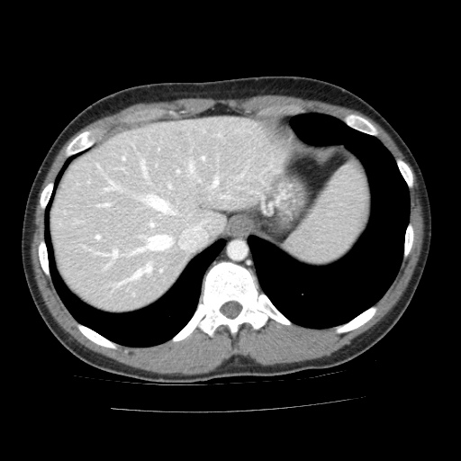
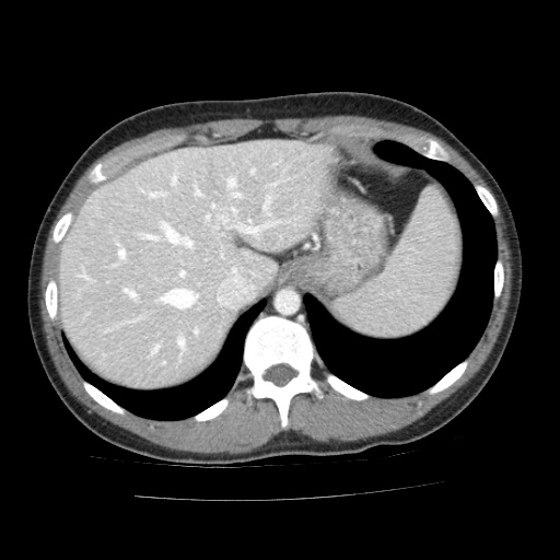

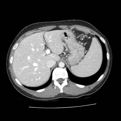
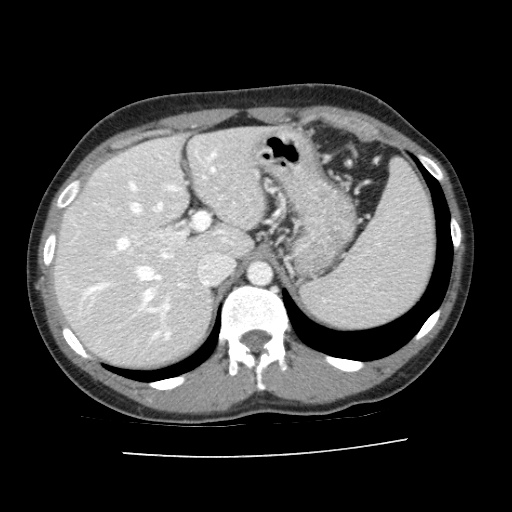
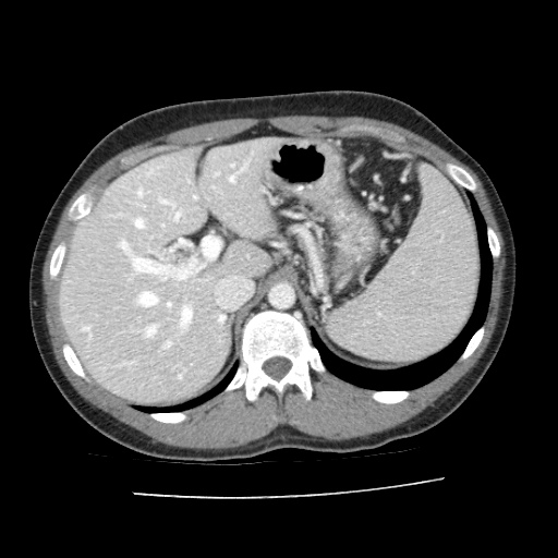
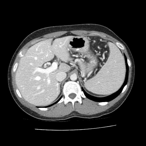


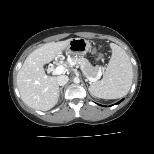
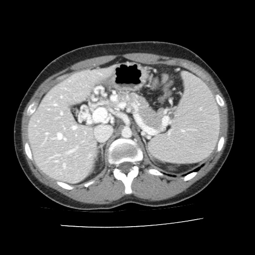
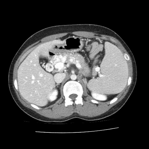
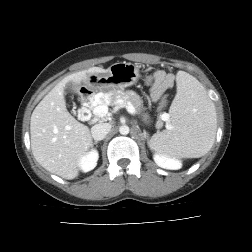

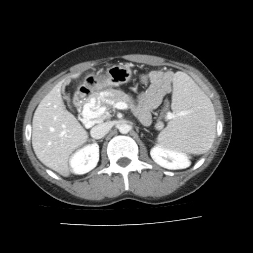
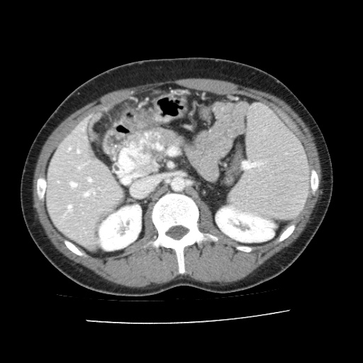
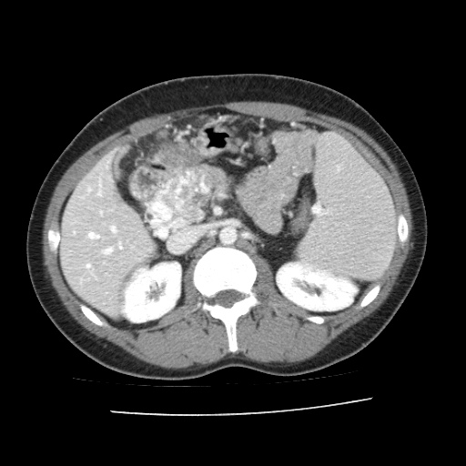

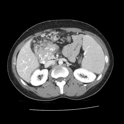
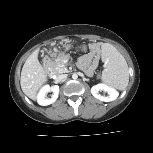
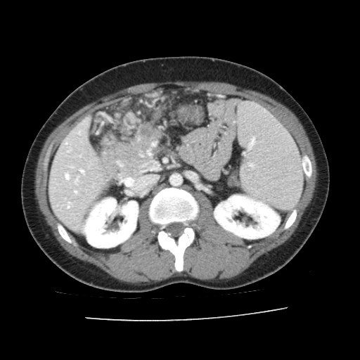

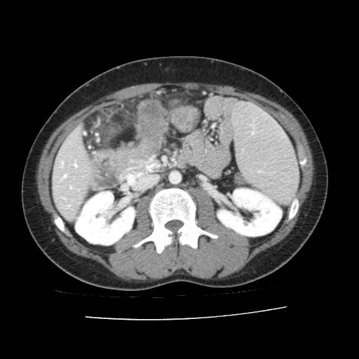
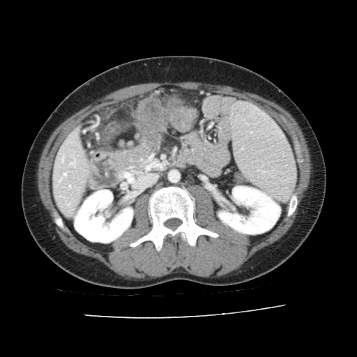

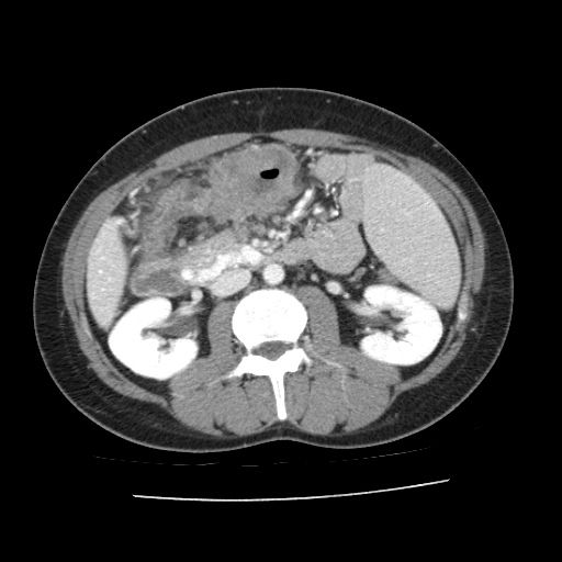
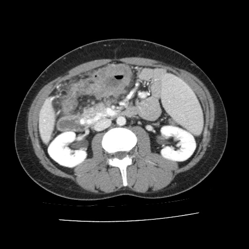
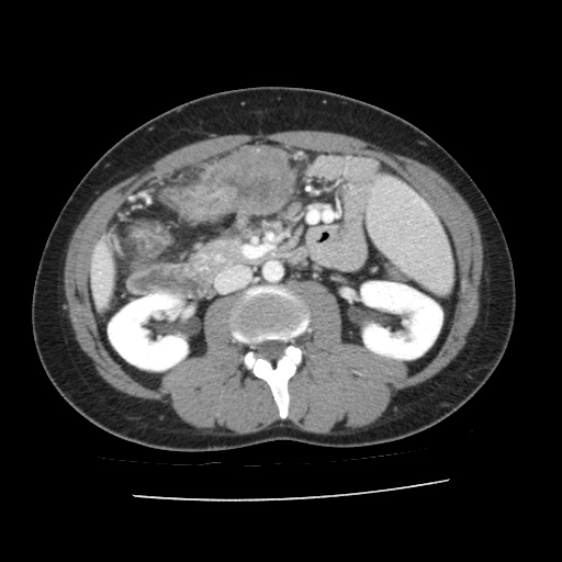
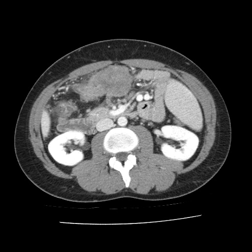
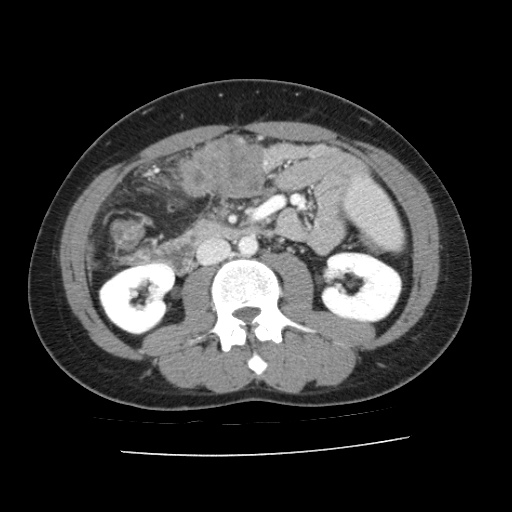
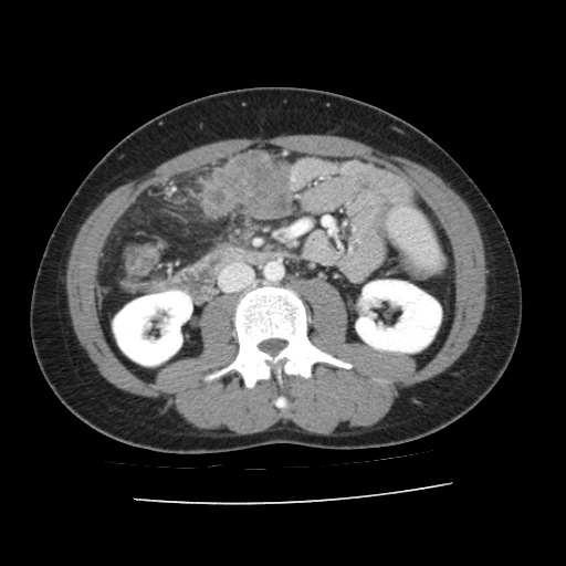

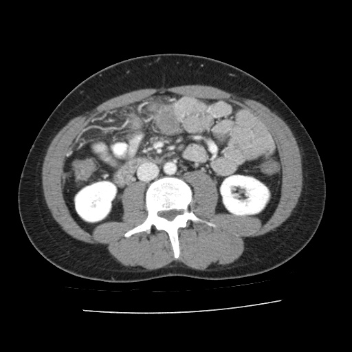

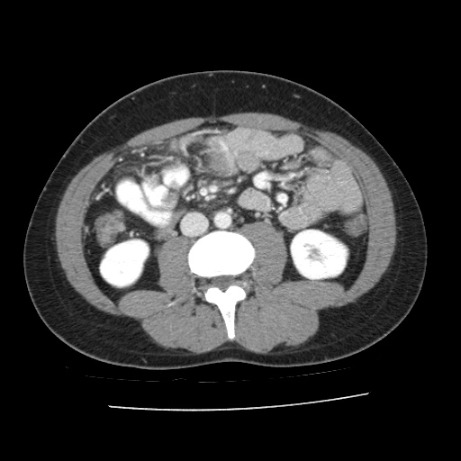
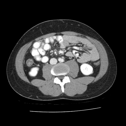
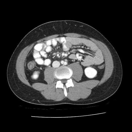
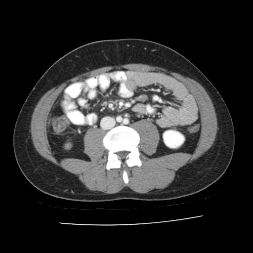
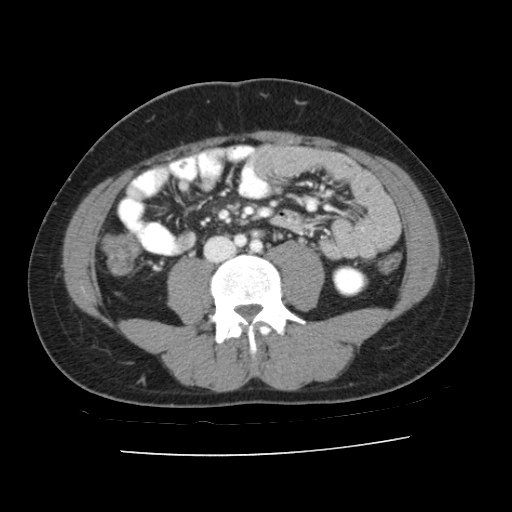
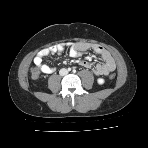


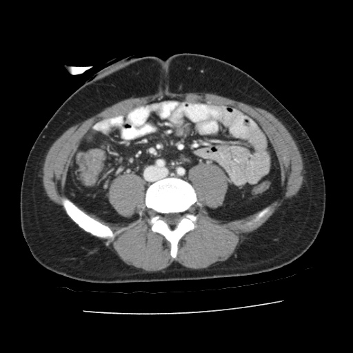
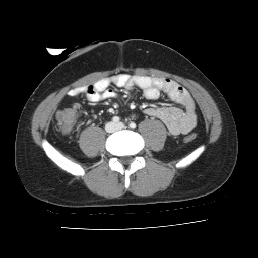


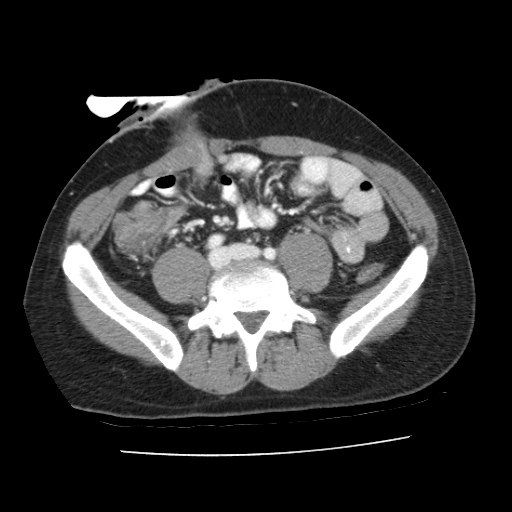
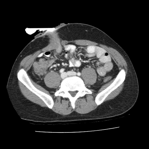

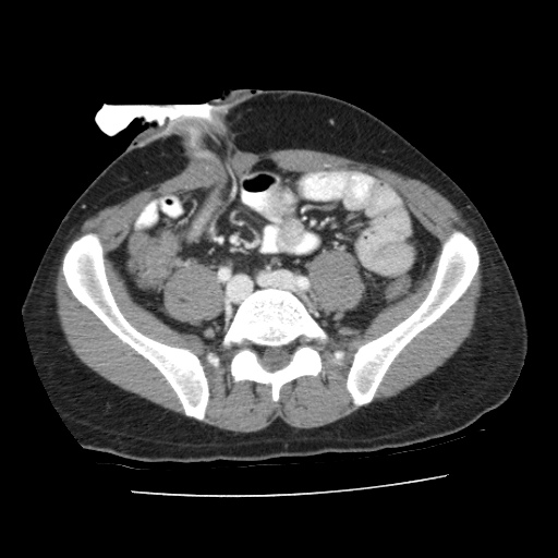


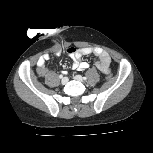
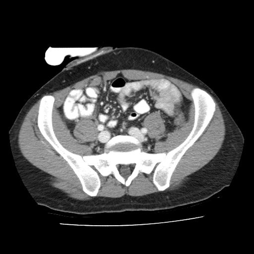


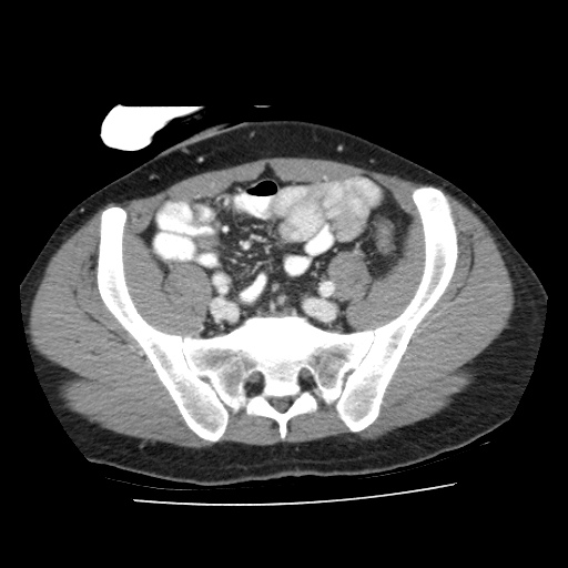

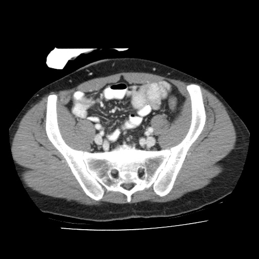
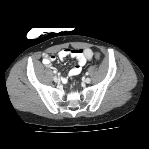
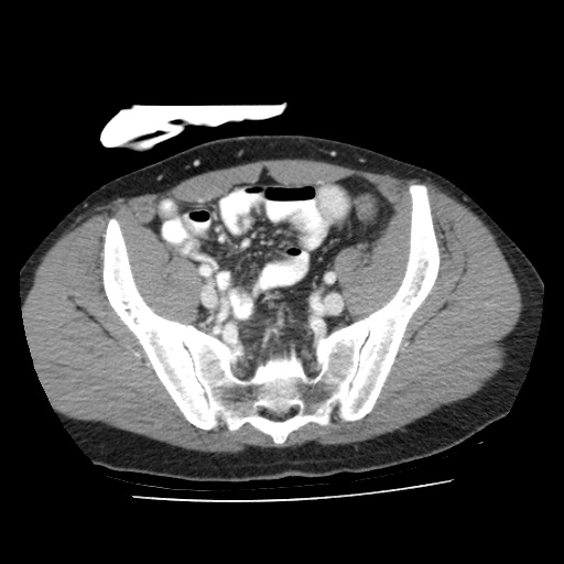

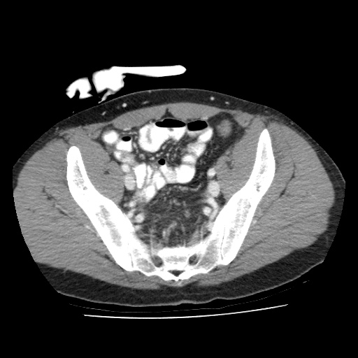
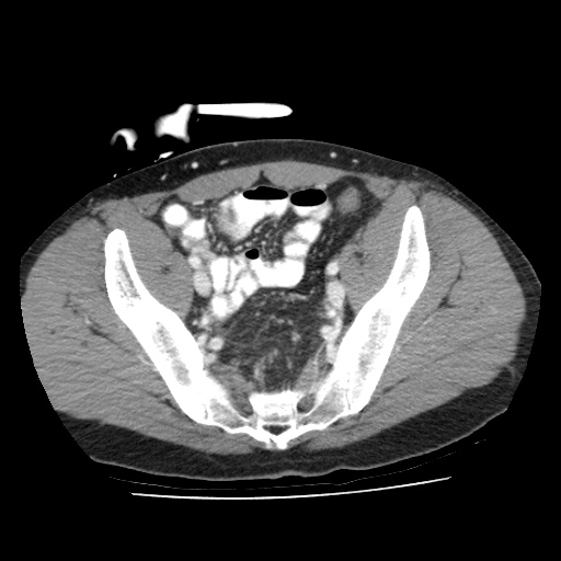
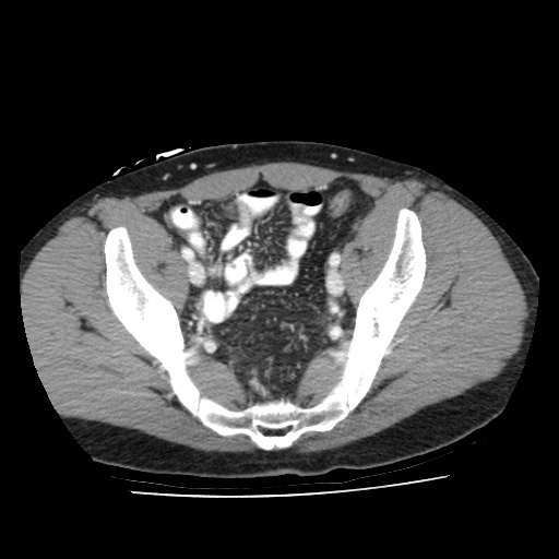
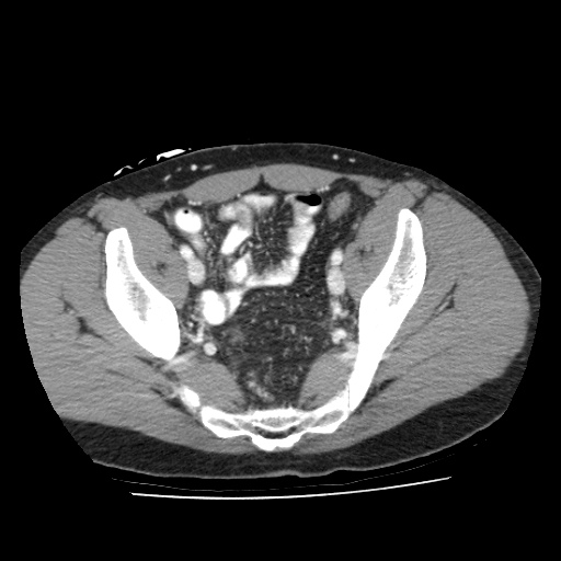
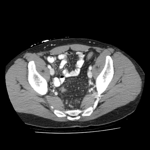
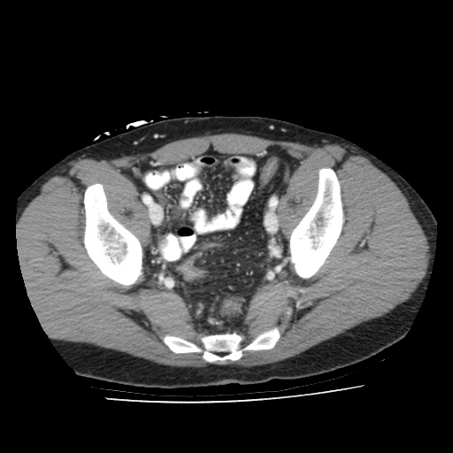
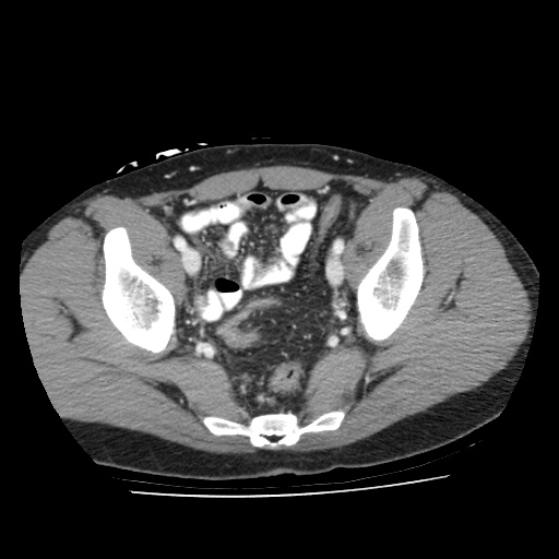
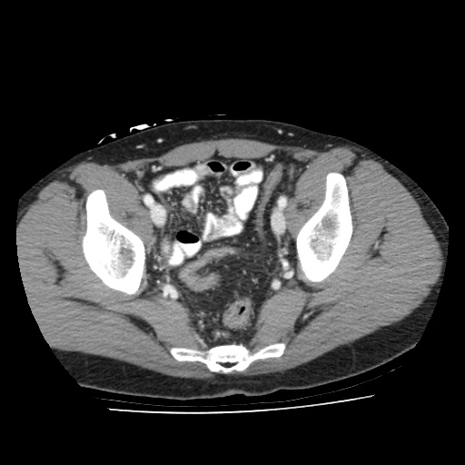
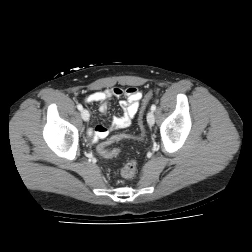

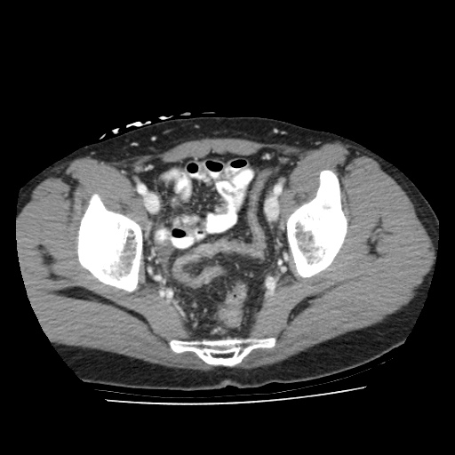


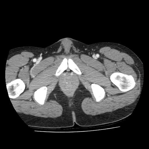


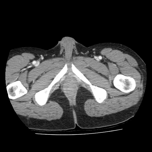
①
Venous collateral branches developed around the pancreas head, as a result of an obstructed SMV.
②
Obstructed SMV shown here. Scroll inferiorly to observe the SMV and its jejunal branches.
③
Obstructive tumor that originated from the transverse colon.
④
1st jejunal branch of the SMV, which will be divided and reconstructed.
Click to turn annotations on/off
- Click and drag the slider on the left to transverse through the venous CT series. Clicking on the grey button on the bottom will reveal clinical findings by the surgical team; click on the descriptions to bring the image up to the main window. Best viewed in Mozilla Firefox, Google Chrome or Safari.
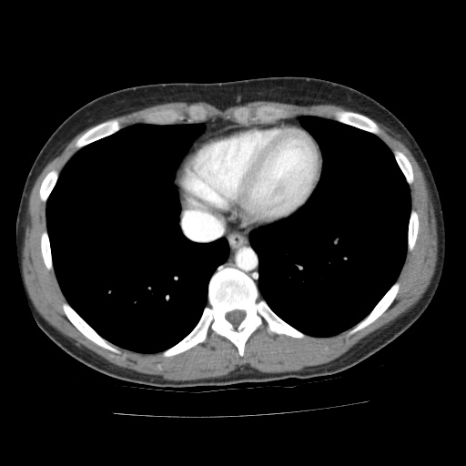
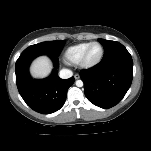
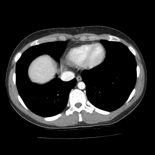
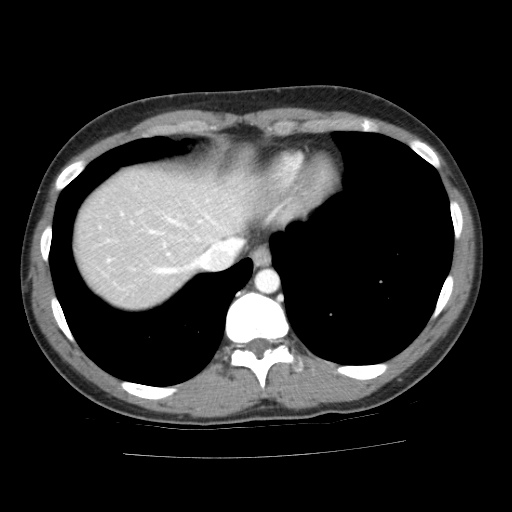
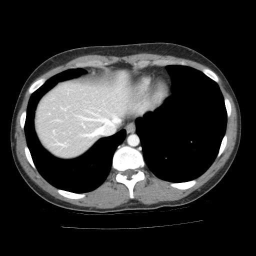
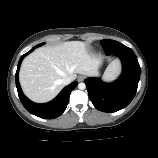
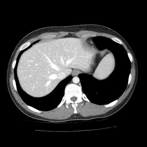
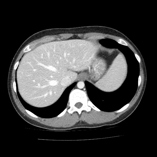
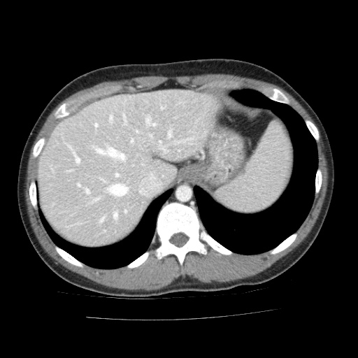
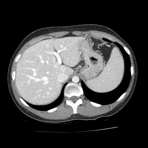
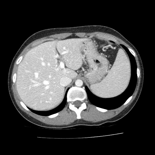
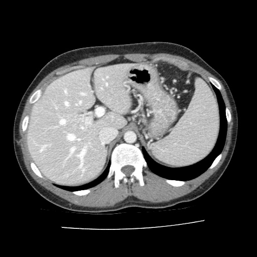
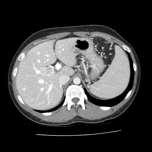
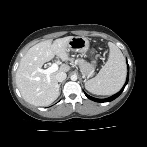
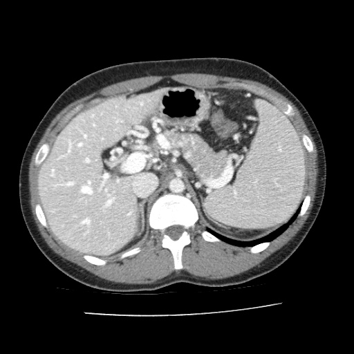
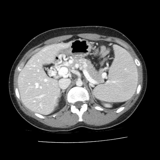
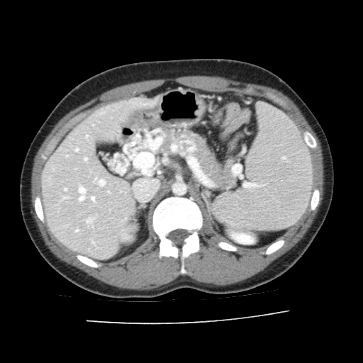
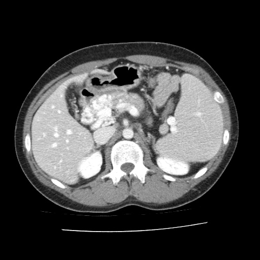
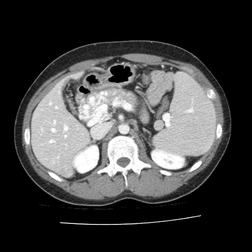
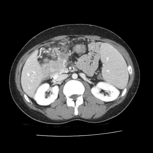
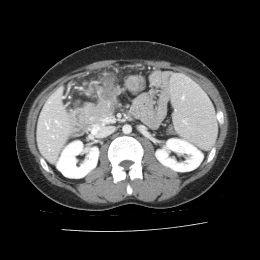
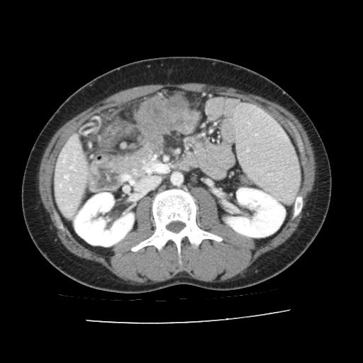
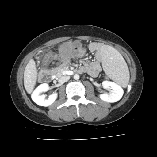
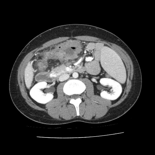
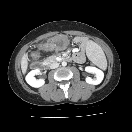
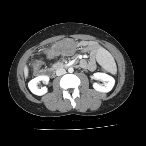
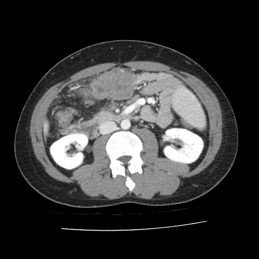
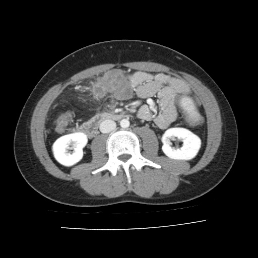
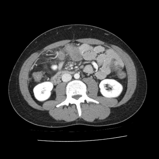
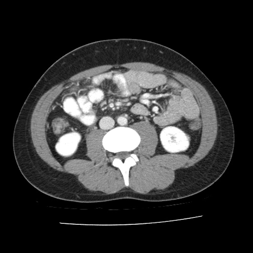
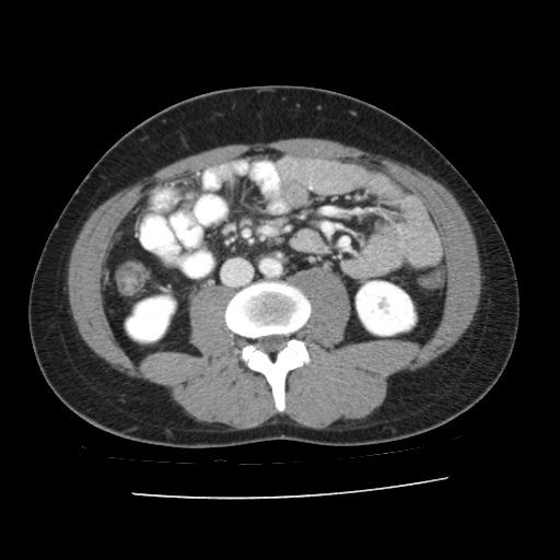
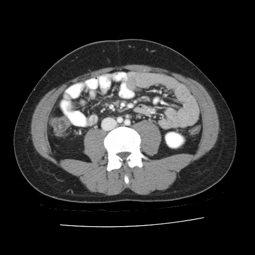
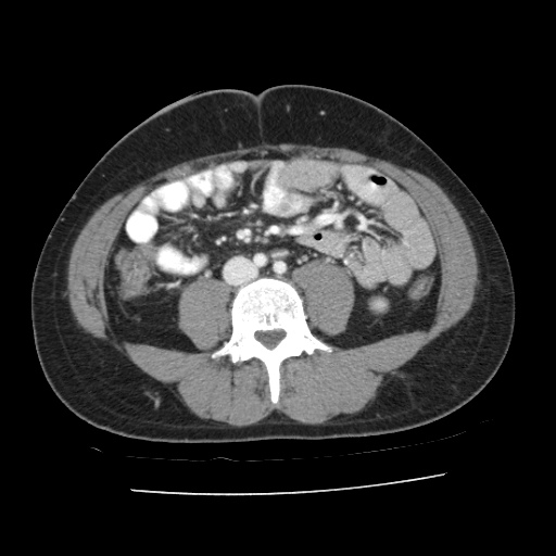
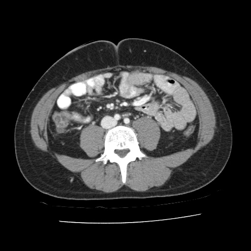
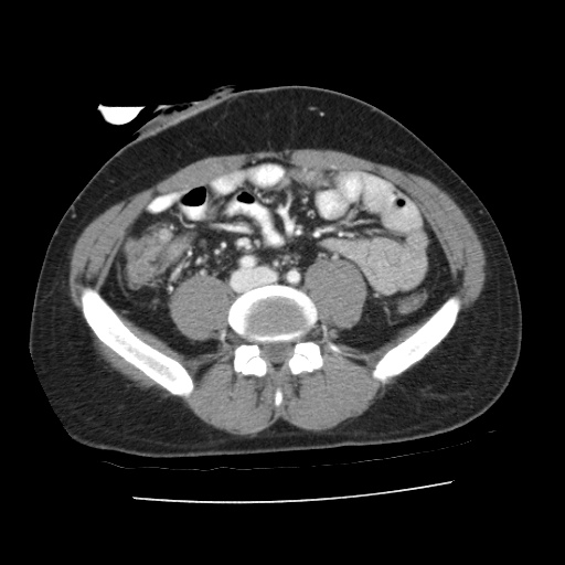
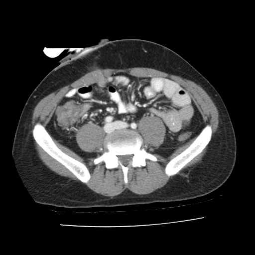
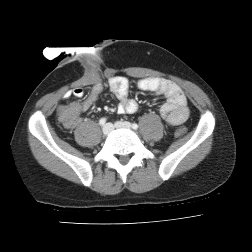
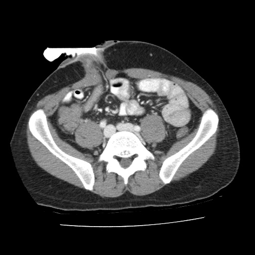
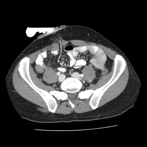
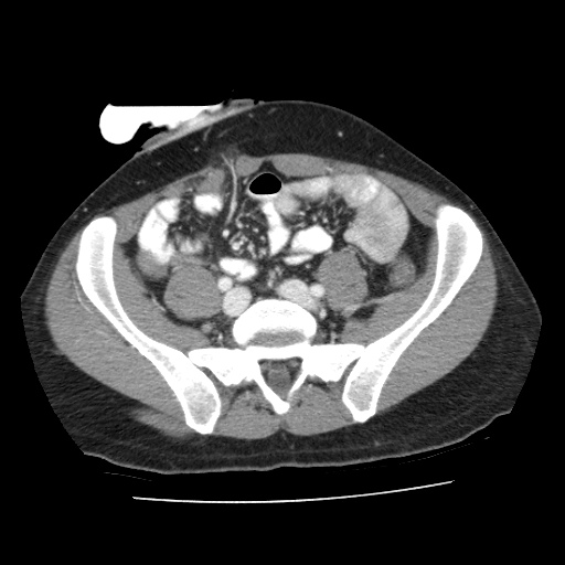
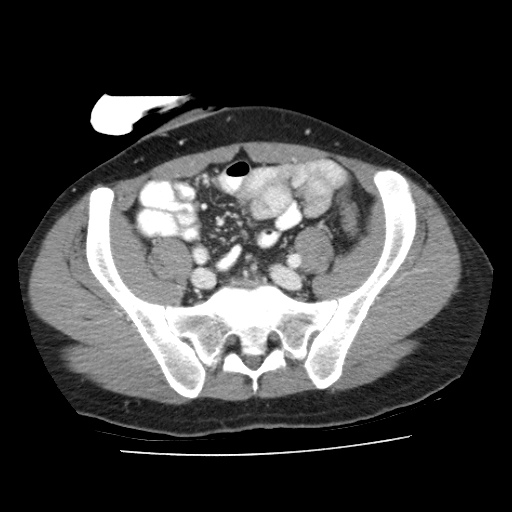
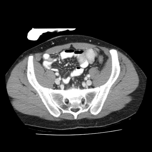
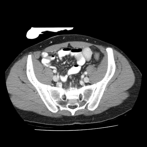
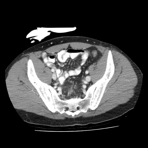
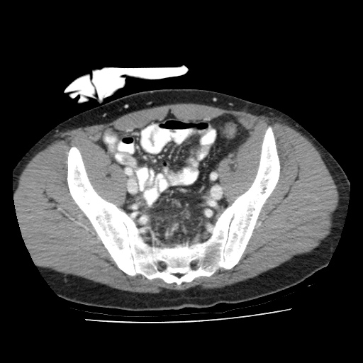
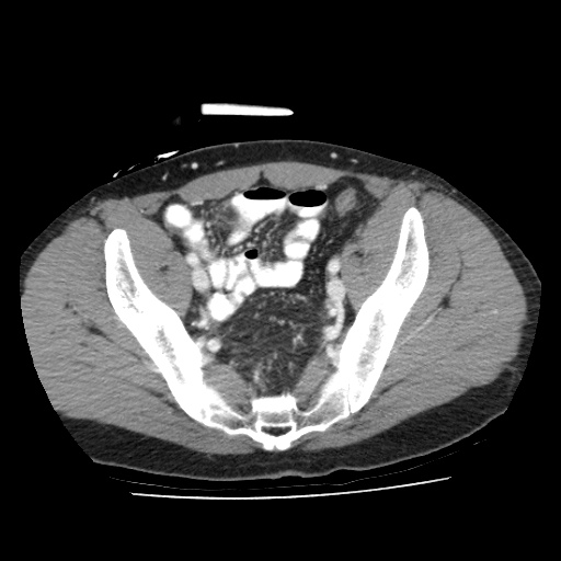
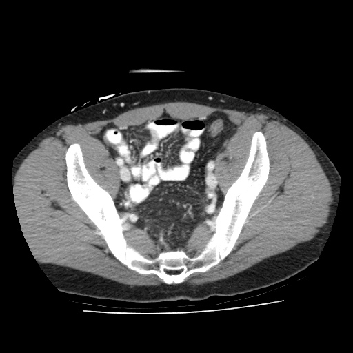
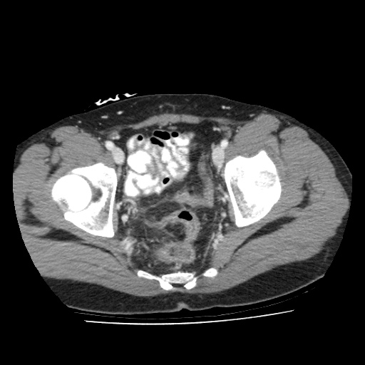
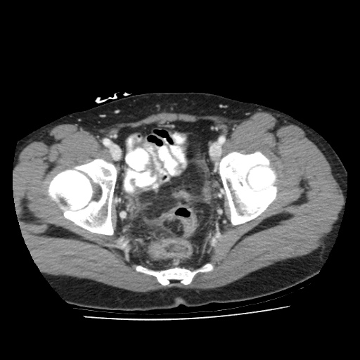
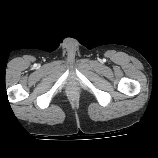
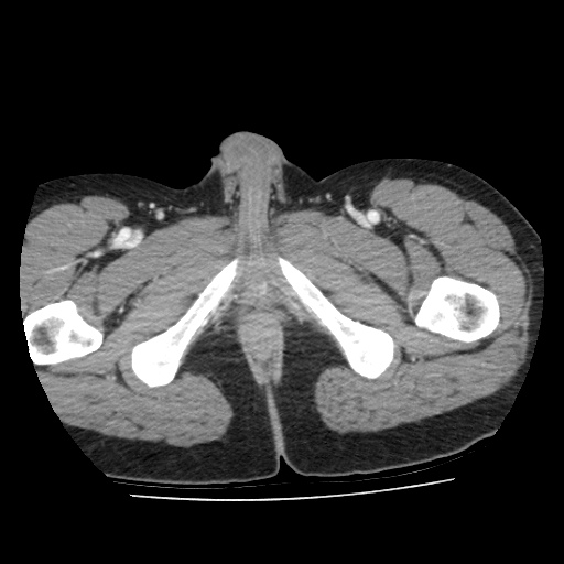
CT scans (arterial phase)
- Click and drag the slider on the left to transverse through the arterial CT series. Clicking on the grey button on the bottom will reveal clinical findings by the surgical team; click on the descriptions to bring the image up to the main window. Best viewed in Mozilla Firefox, Google Chrome or Safari.


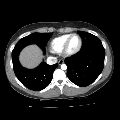
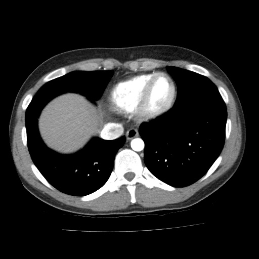


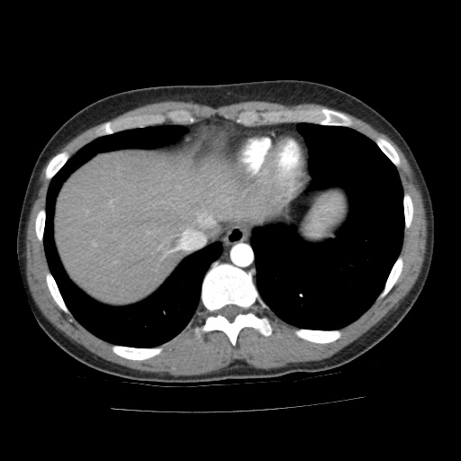
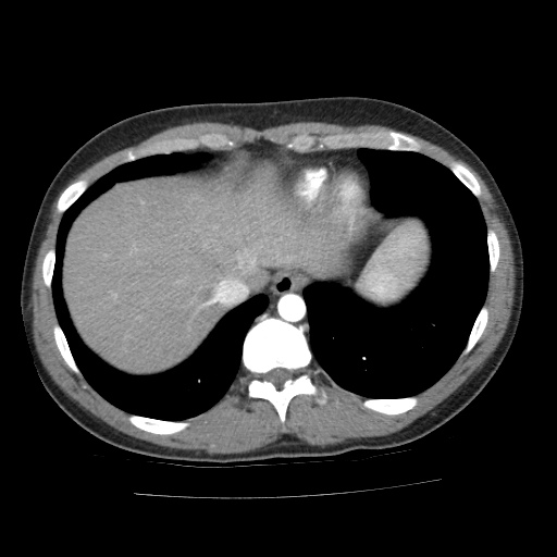
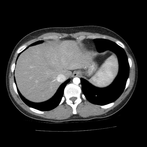
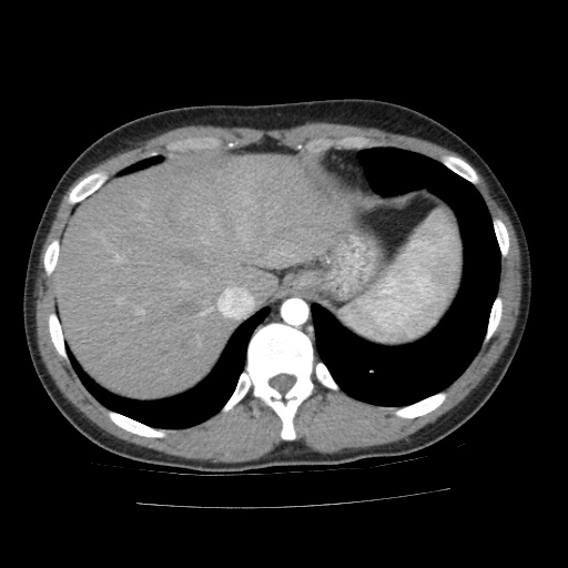

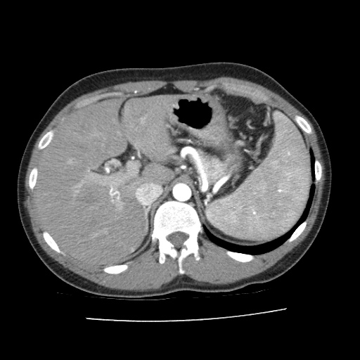
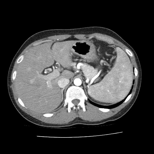
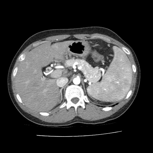
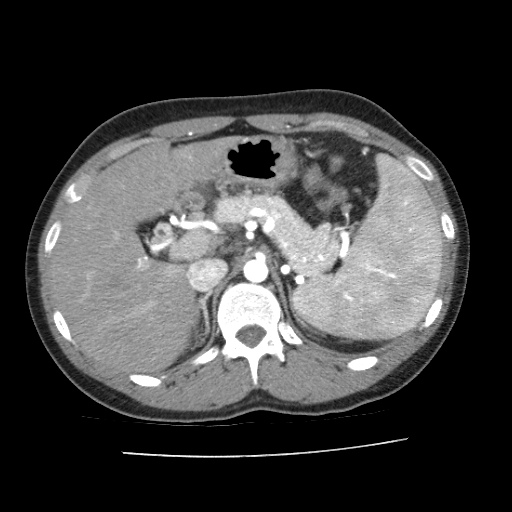

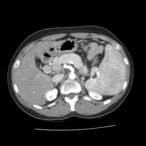
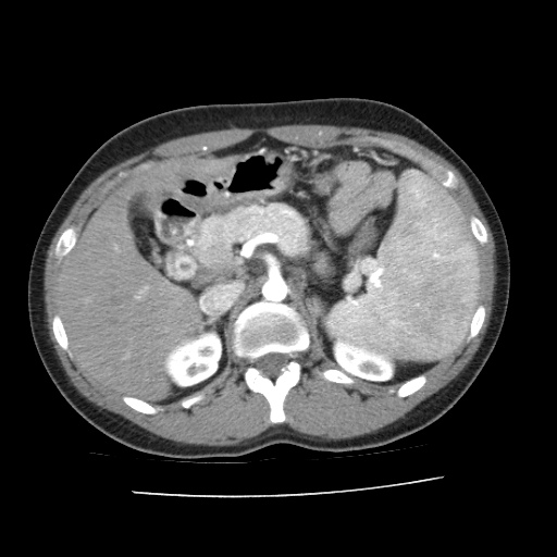

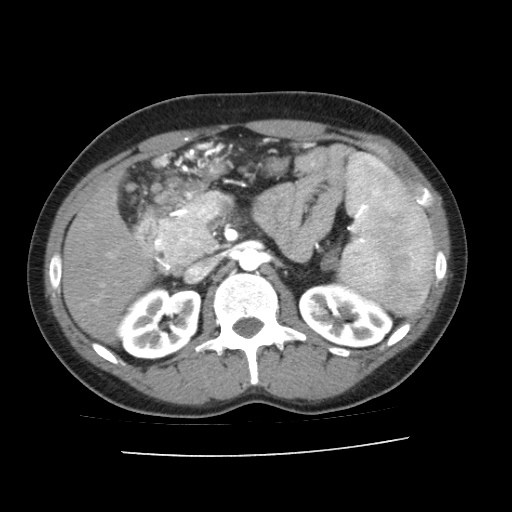

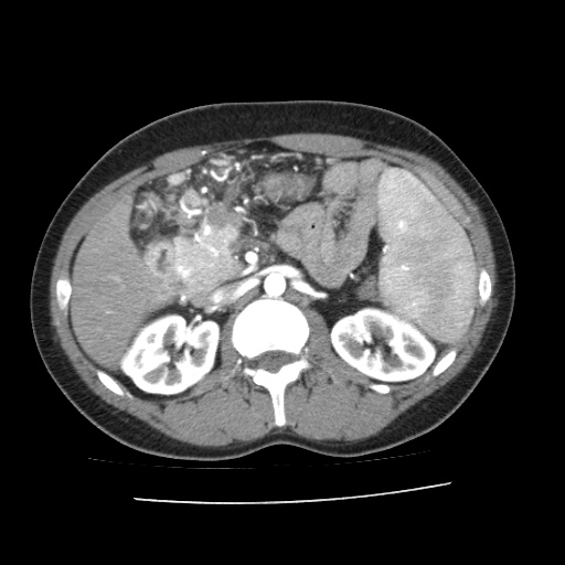
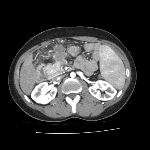
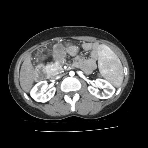


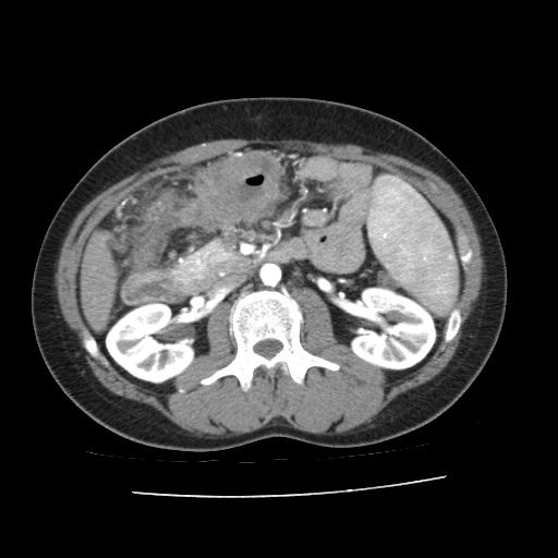
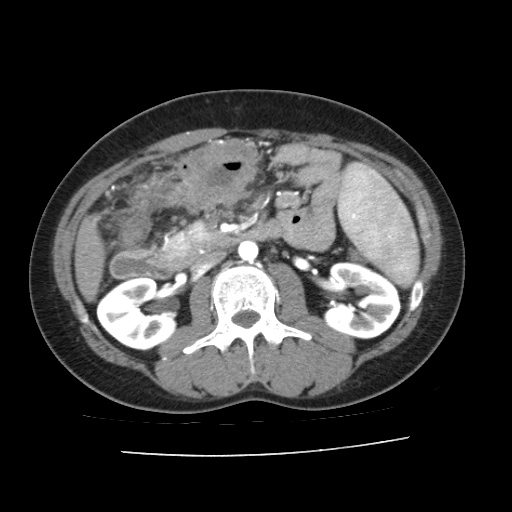
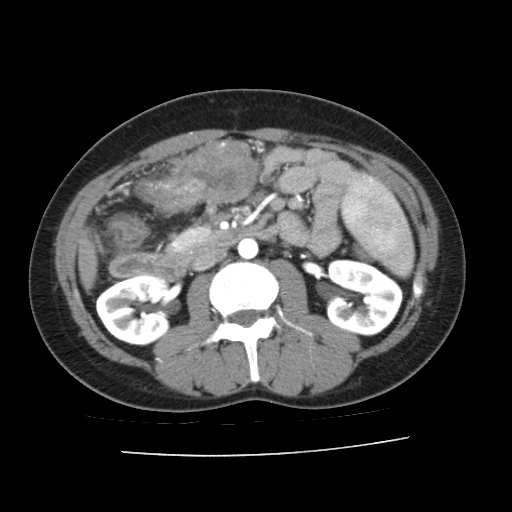
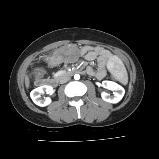
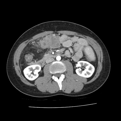
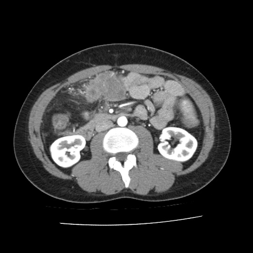
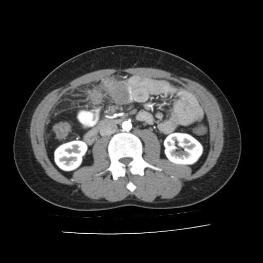
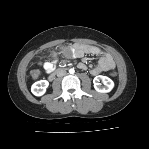
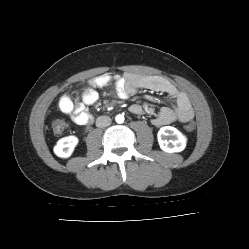
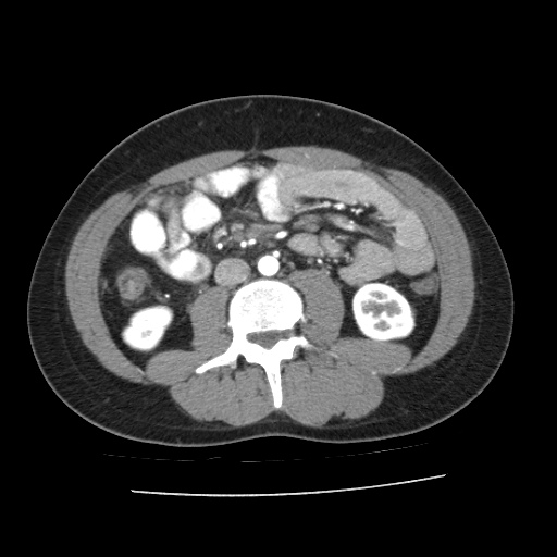

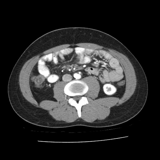
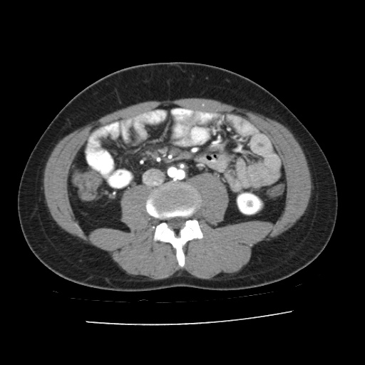

①
Obstructive tumor and SMA shown here. The tumor is shown abutting the SMA.
②
Jejunal branches of the SMA shown here. These branches will be preserved.
Click to turn annotations on/off
- Click and drag the slider on the left to transverse through the arterial CT series. Clicking on the grey button on the bottom will reveal clinical findings by the surgical team; click on the descriptions to bring the image up to the main window. Best viewed in Mozilla Firefox, Google Chrome or Safari.
Related cases