Klatskin tumours: Extended left hepatectomy
With complex portal vein reconstruction and in situ cold perfusion of the liver
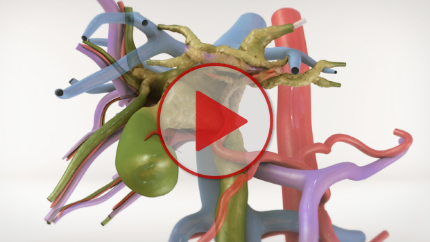
View the Video
HD
is off
Video Chapters


Drag slider to navigate through video chapters.
Case Description
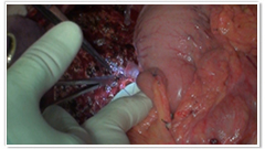
- The patient was a 42-year-old male with a locally advanced Klatskin tumour.
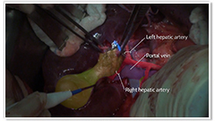
- The tumour originated at the left margin of the hepatic duct bifurcation. It invaded the anterior branch of the right hepatic artery and the left hepatic artery. It also invaded the left portal vein and the anterior branch of the right portal vein. However, an accessory segment 7 and segment 6/7 branches of the right portal vein were free of tumour.
- To optimize the liver for surgery, biliary system drainage and left PV embolization were performed.
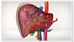
- An extended left hepatectomy involving segments 1 to 5 and 8 was planned, preserving segments 6 and 7.
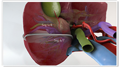
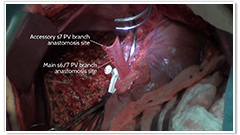
- This transection line divided both an accessory segment 7 and segment 6/7 branches of the right portal vein. Both were reconstructed to the main portal vein during in-situ cold perfusion.
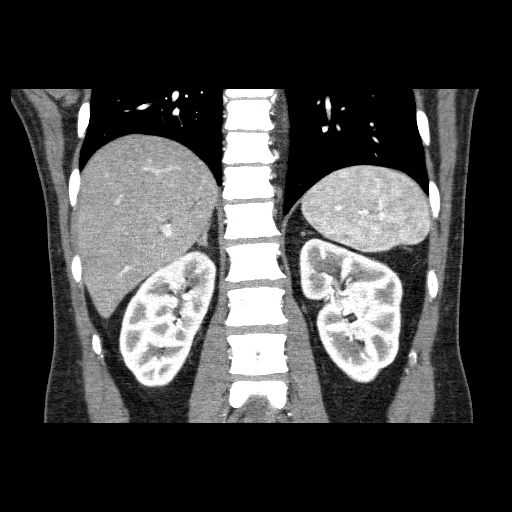
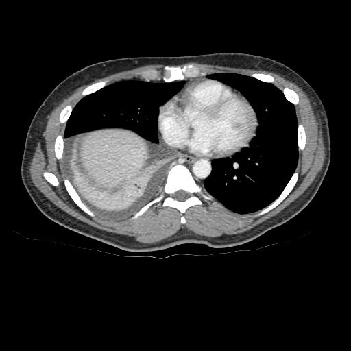
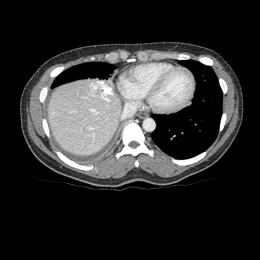
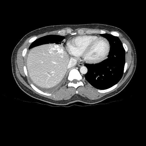
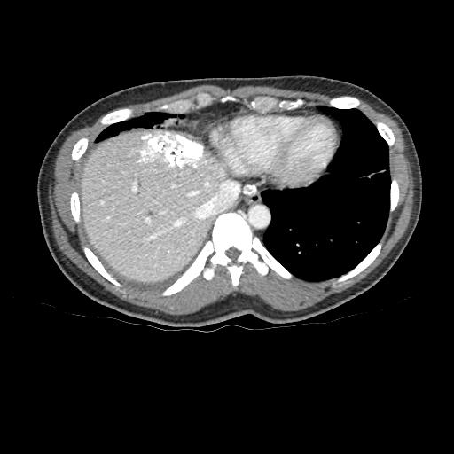
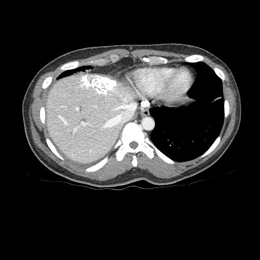
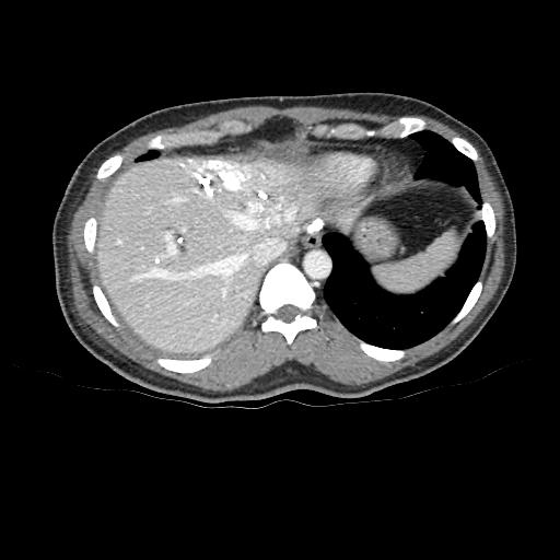
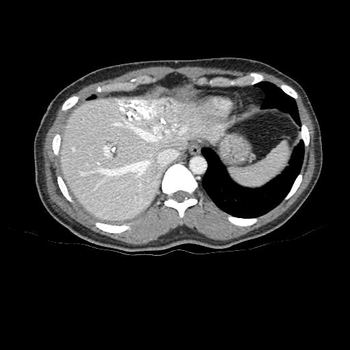
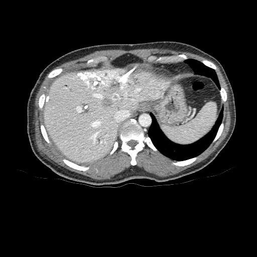
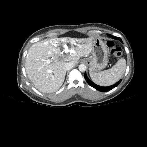
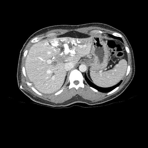
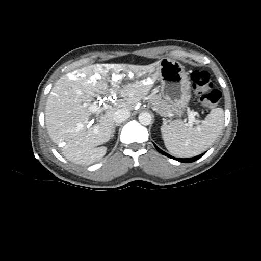
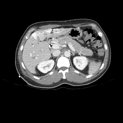
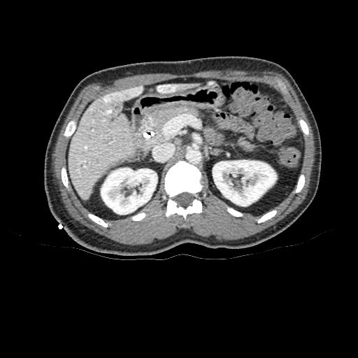
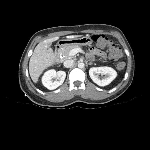
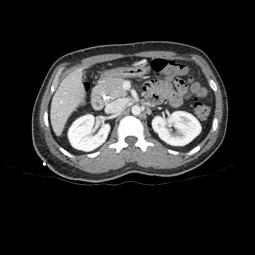
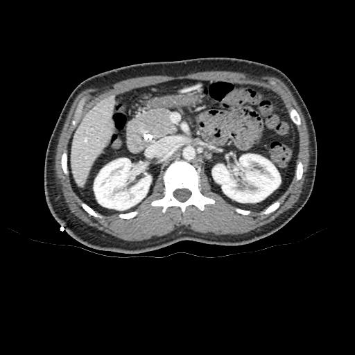
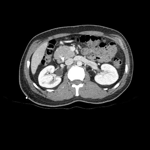
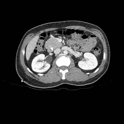
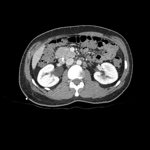
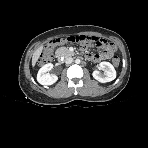
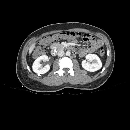
CT scans (Pre-portal vein embolization)
- Click and drag the slider on the left to transverse through the CT series. Clicking on the grey button on the bottom will reveal clinical findings by the surgical team; click on the descriptions to bring the image up to the main window. Best viewed in Mozilla Firefox, Google Chrome or Safari.
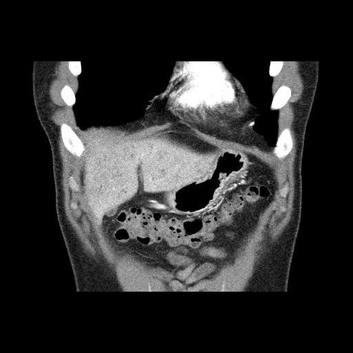
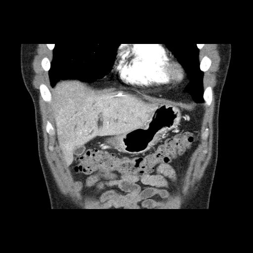
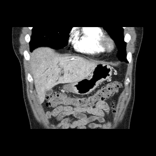
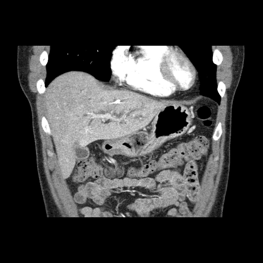
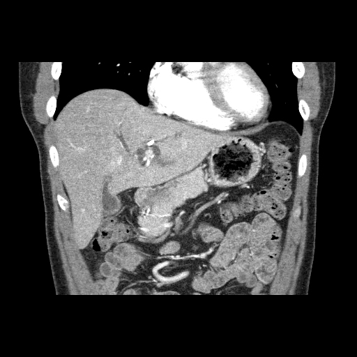
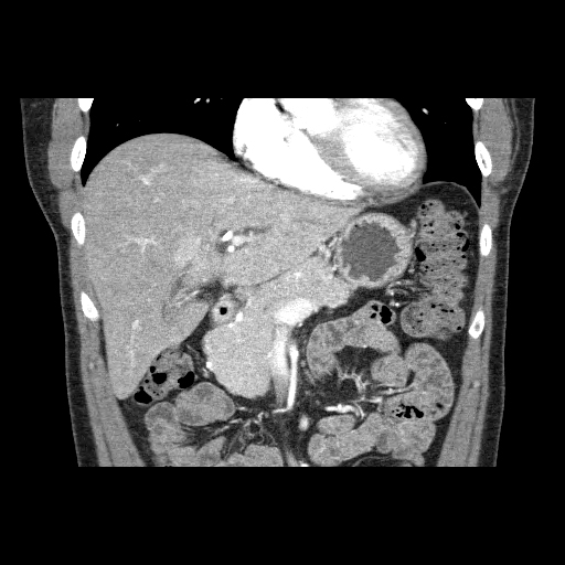
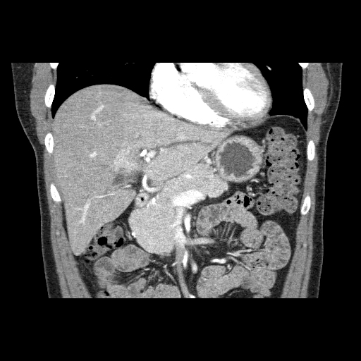


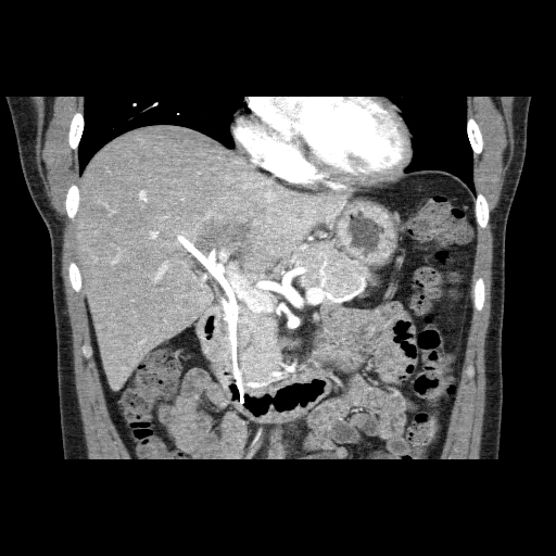

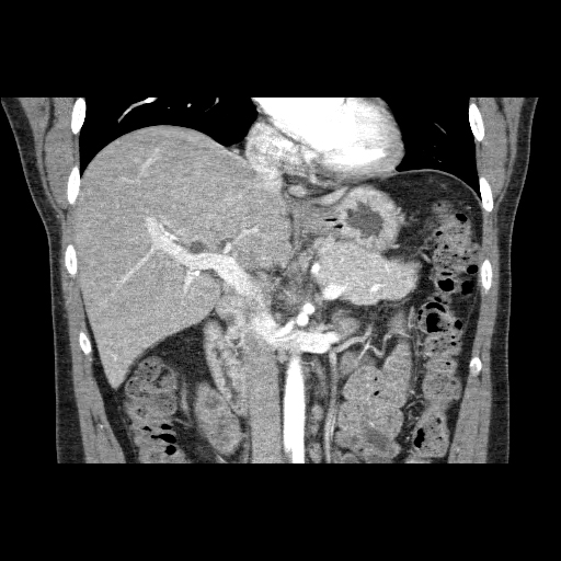
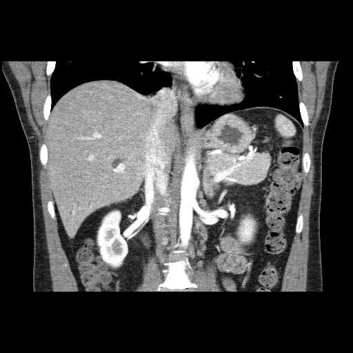
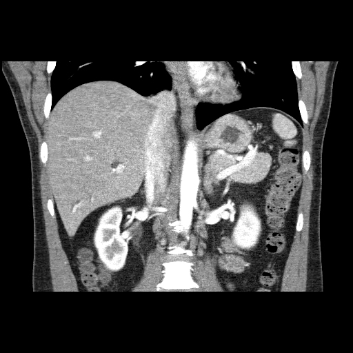
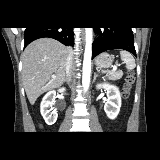

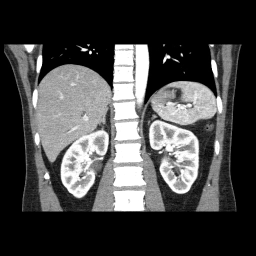

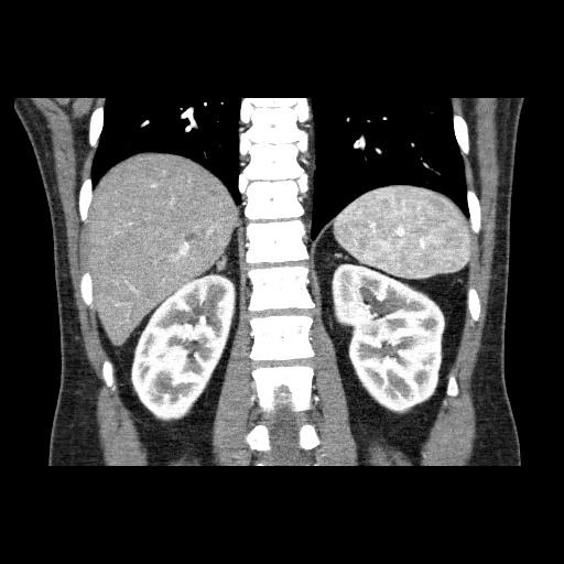
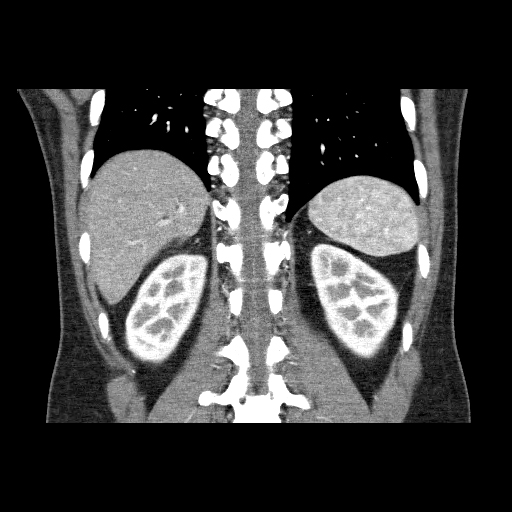

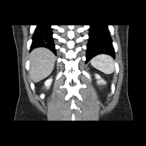
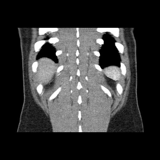
①
Acessory segment 7 and main segment 6/7 PV branch
Click to turn annotations on/off
- Click and drag the slider on the left to transverse through the CT series. Clicking on the grey button on the bottom will reveal clinical findings by the surgical team; click on the descriptions to bring the image up to the main window. Best viewed in Mozilla Firefox, Google Chrome or Safari.
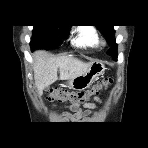
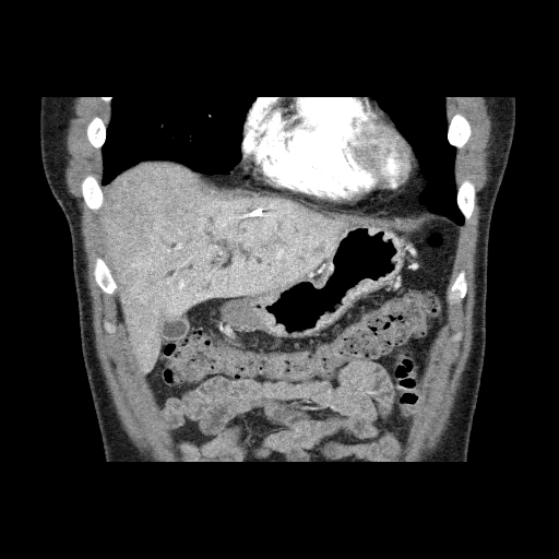
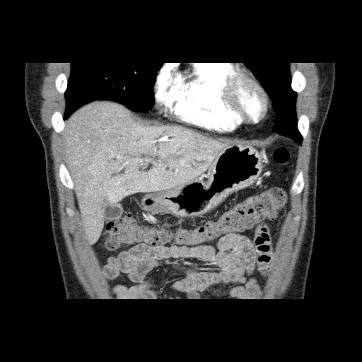
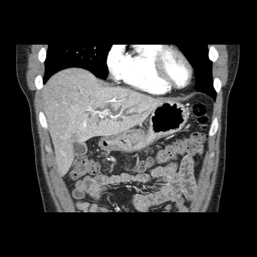
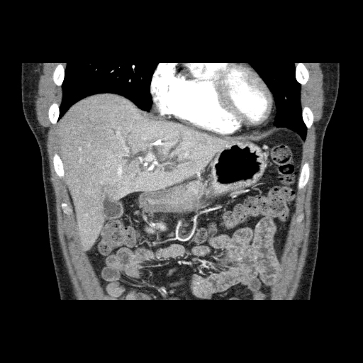
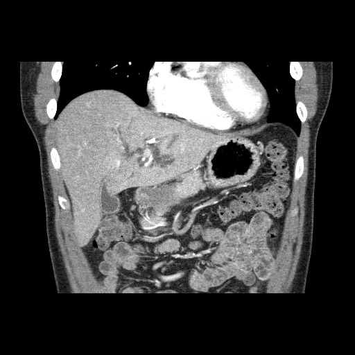
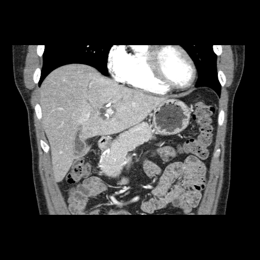
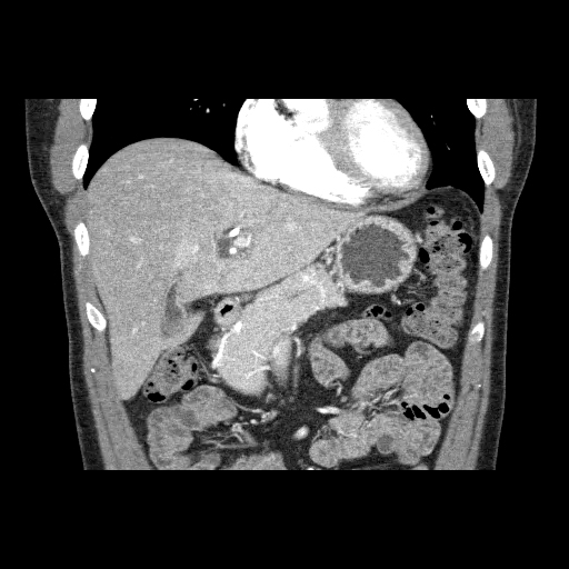
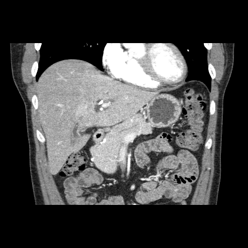
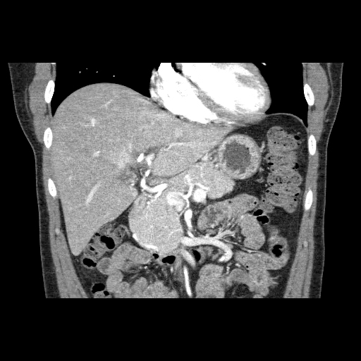
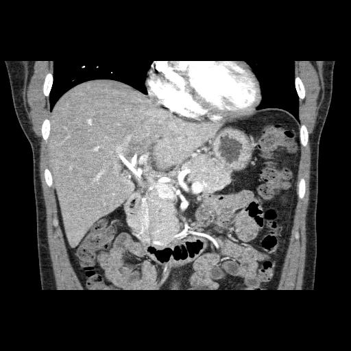
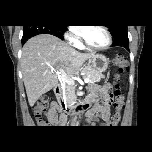
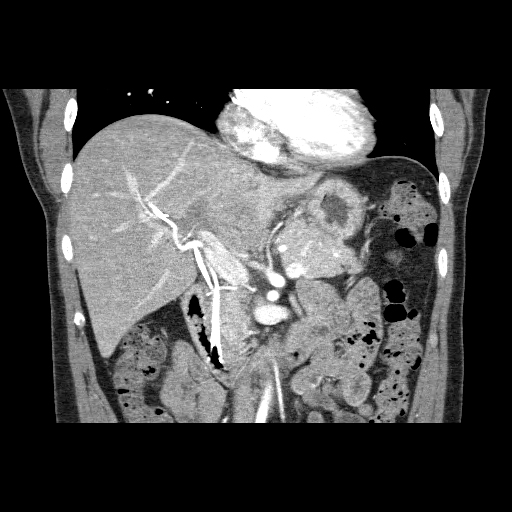
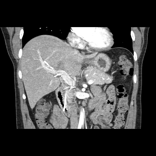
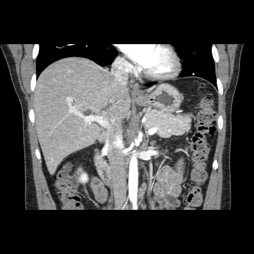
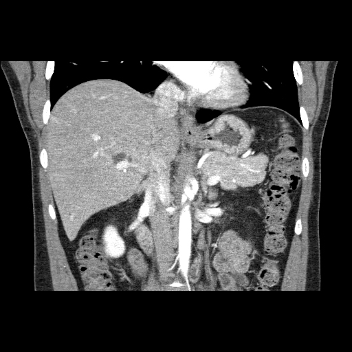
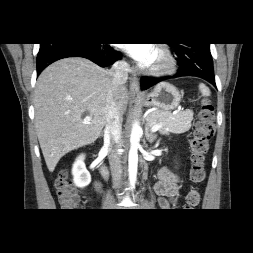
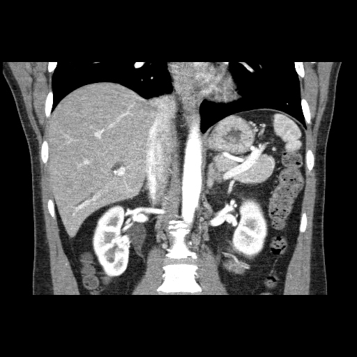
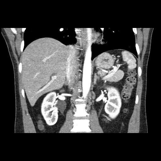
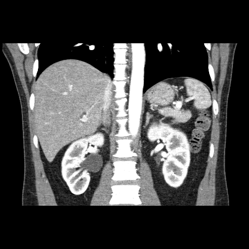
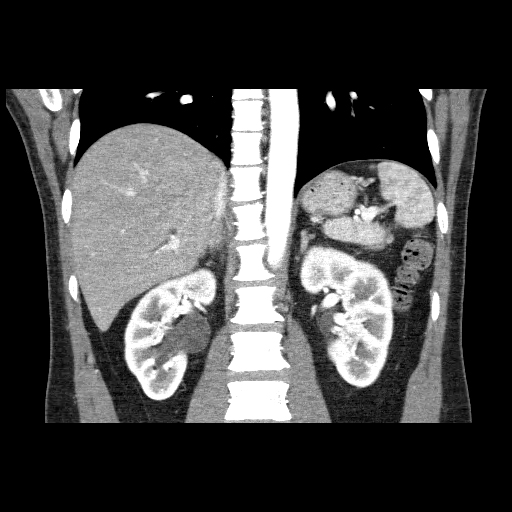
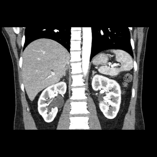
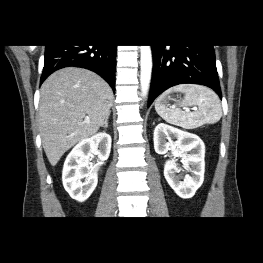
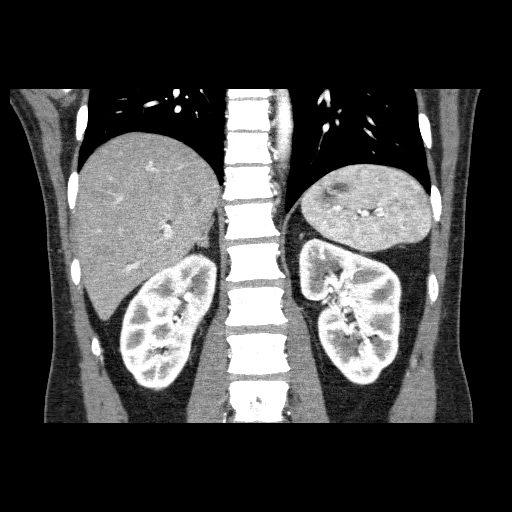
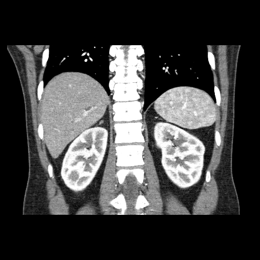
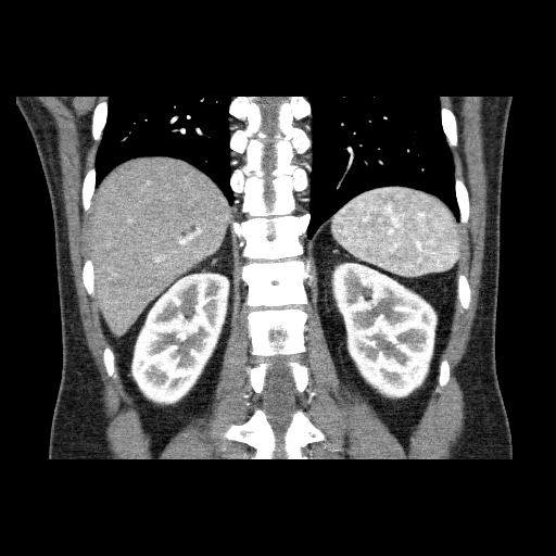
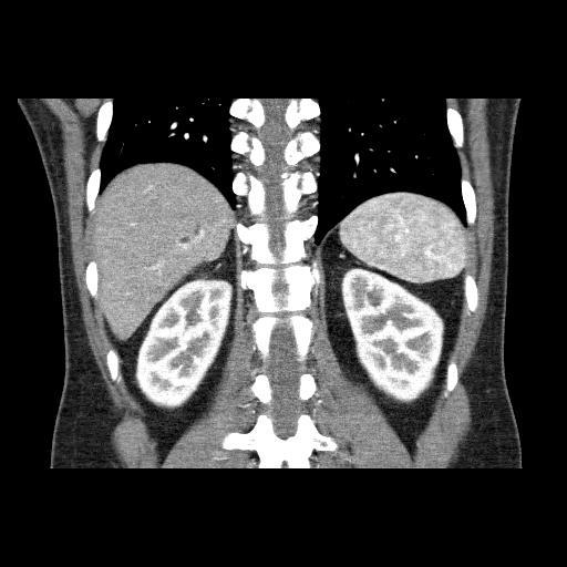
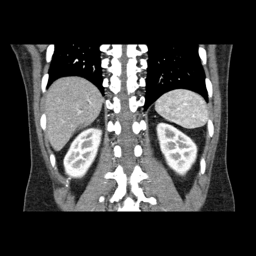
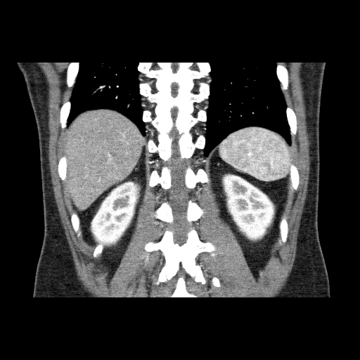
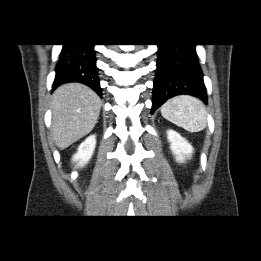
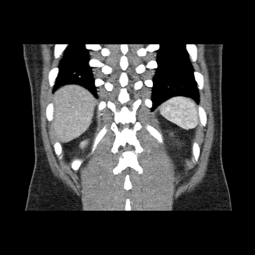
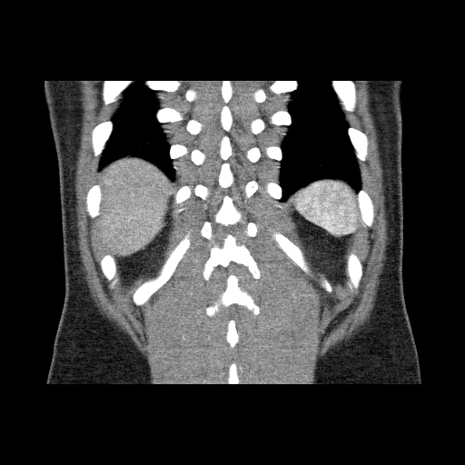
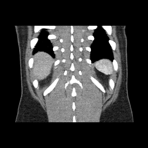
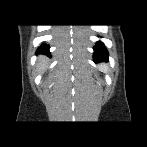
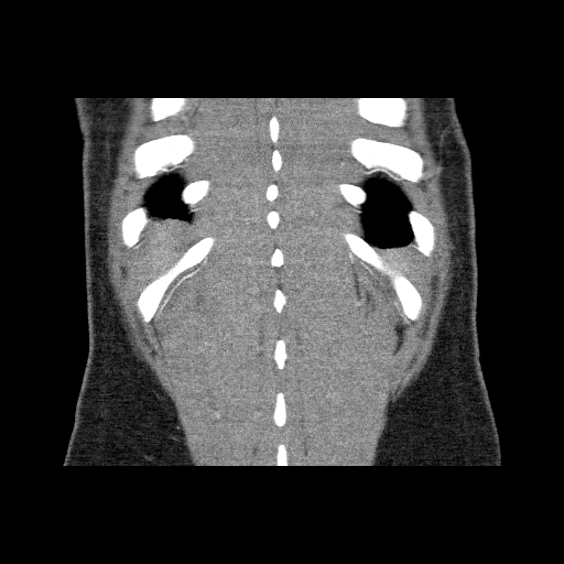
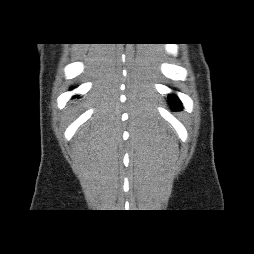
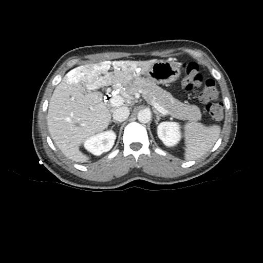
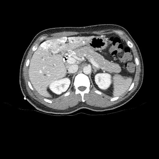
CT scans (Post-portal vein embolization)
- Click and drag the slider on the left to transverse through the CT series. Clicking on the grey button on the bottom will reveal clinical findings by the surgical team; click on the descriptions to bring the image up to the main window. Best viewed in Mozilla Firefox, Google Chrome or Safari.

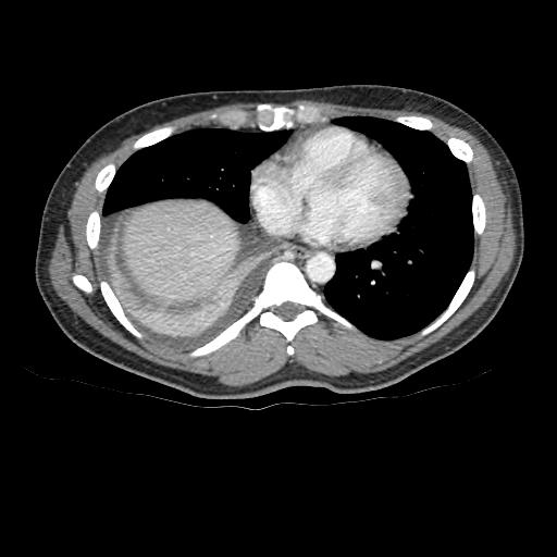
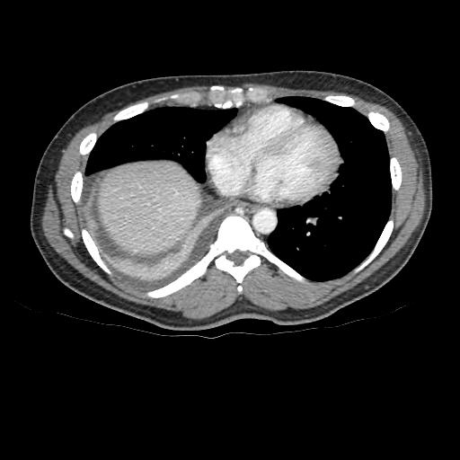
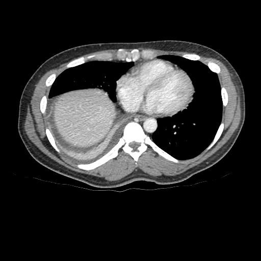


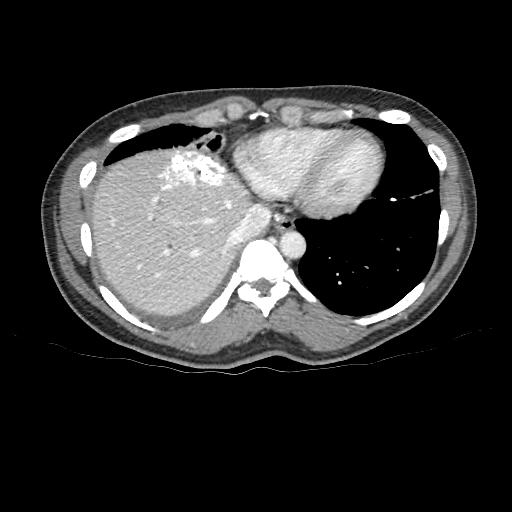
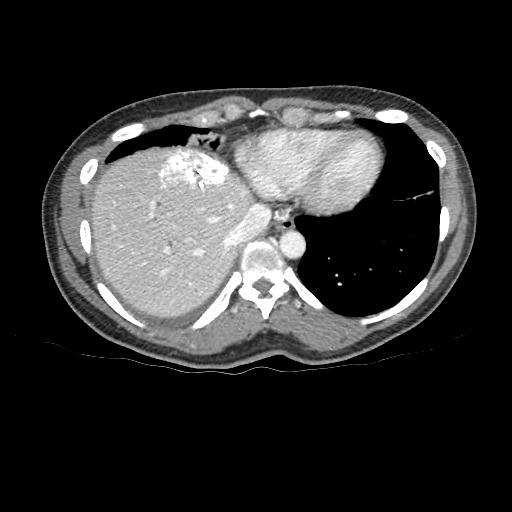


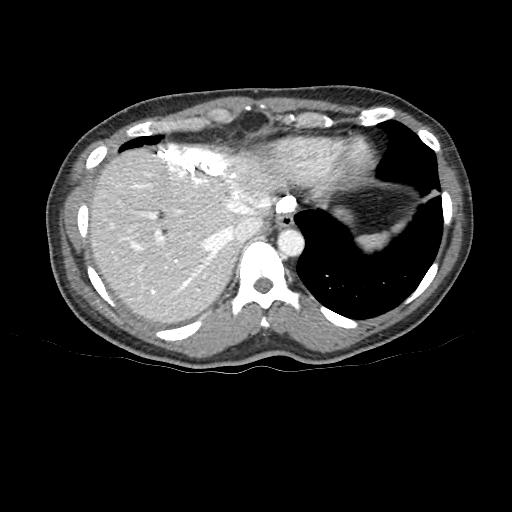
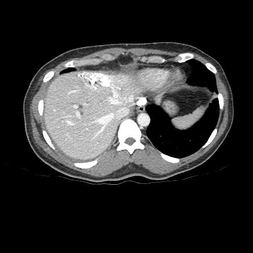
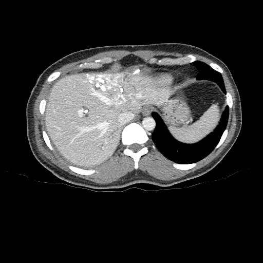
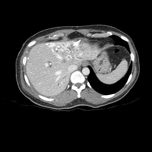
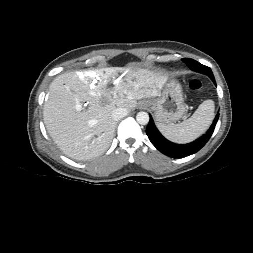
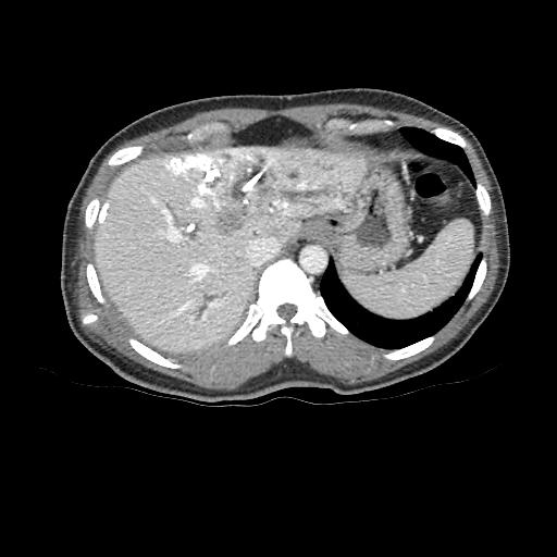

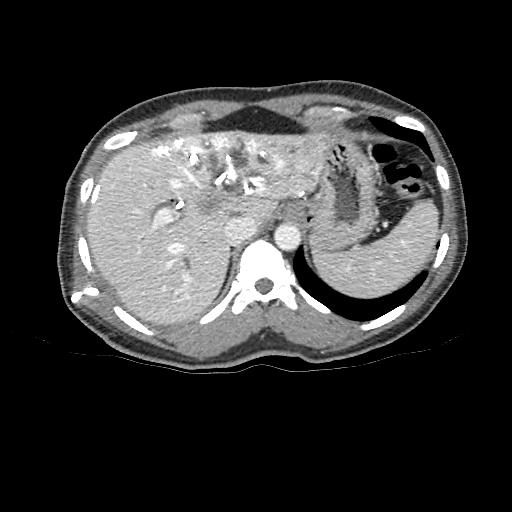
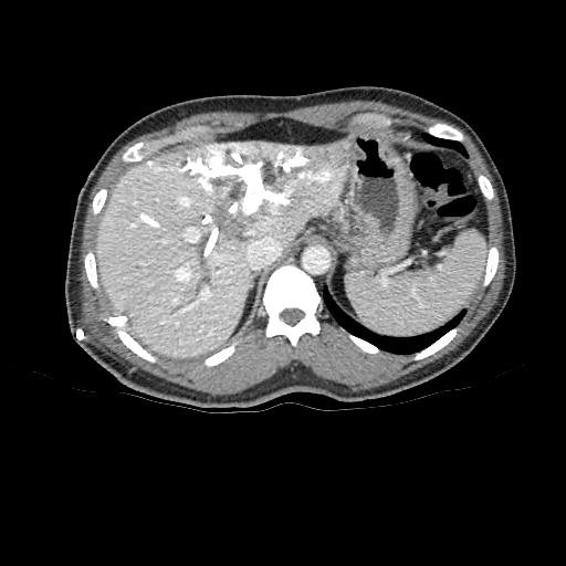
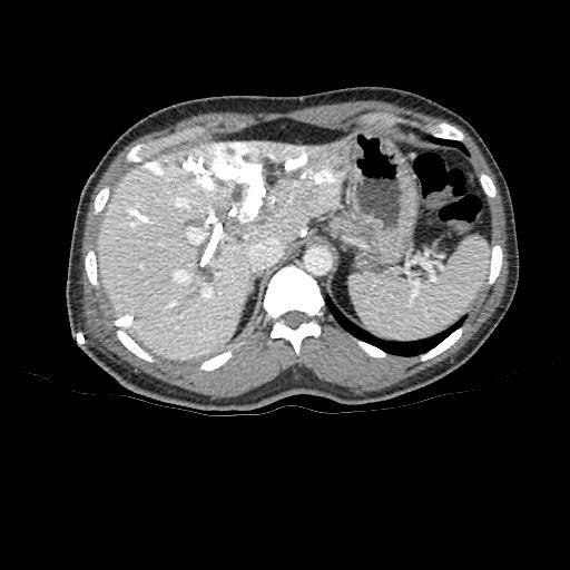


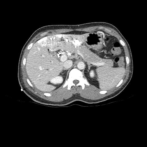


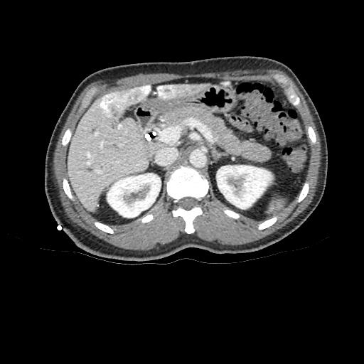
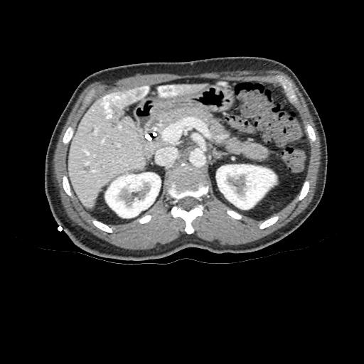
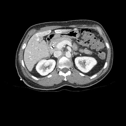
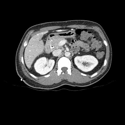
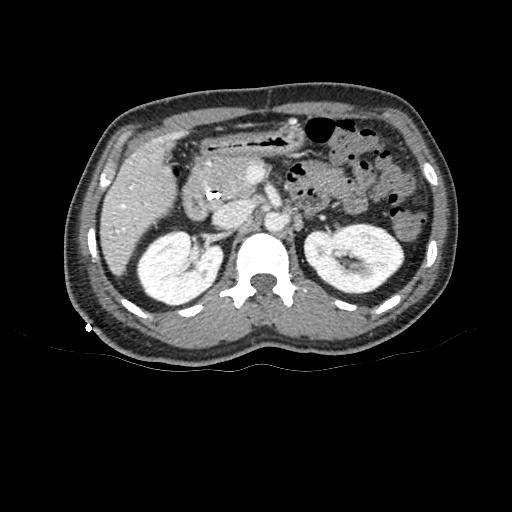

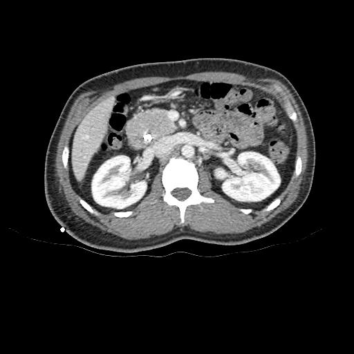
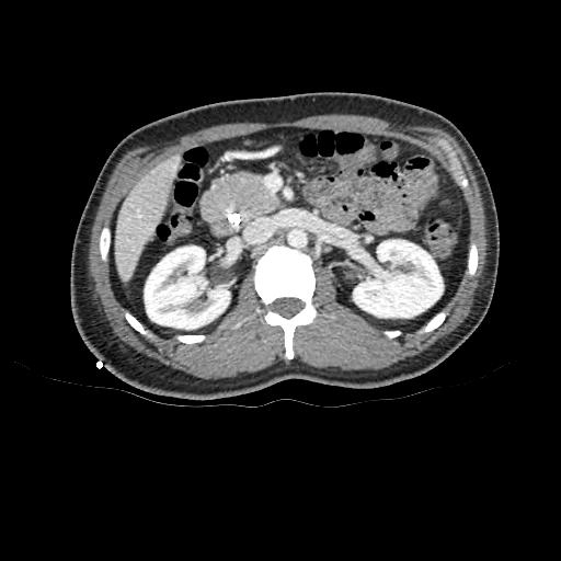
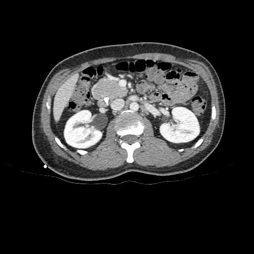
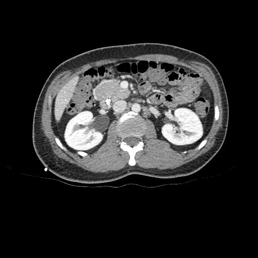
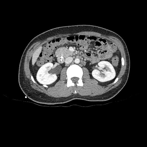
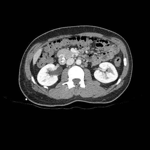
①
MHV is in close proximity to the tumour
②
Accessory segment 7 PV branch
③
Main segment 6/7 PV branch
Click to turn annotations on/off
- Click and drag the slider on the left to transverse through the CT series. Clicking on the grey button on the bottom will reveal clinical findings by the surgical team; click on the descriptions to bring the image up to the main window. Best viewed in Mozilla Firefox, Google Chrome or Safari.